Fig. 1.
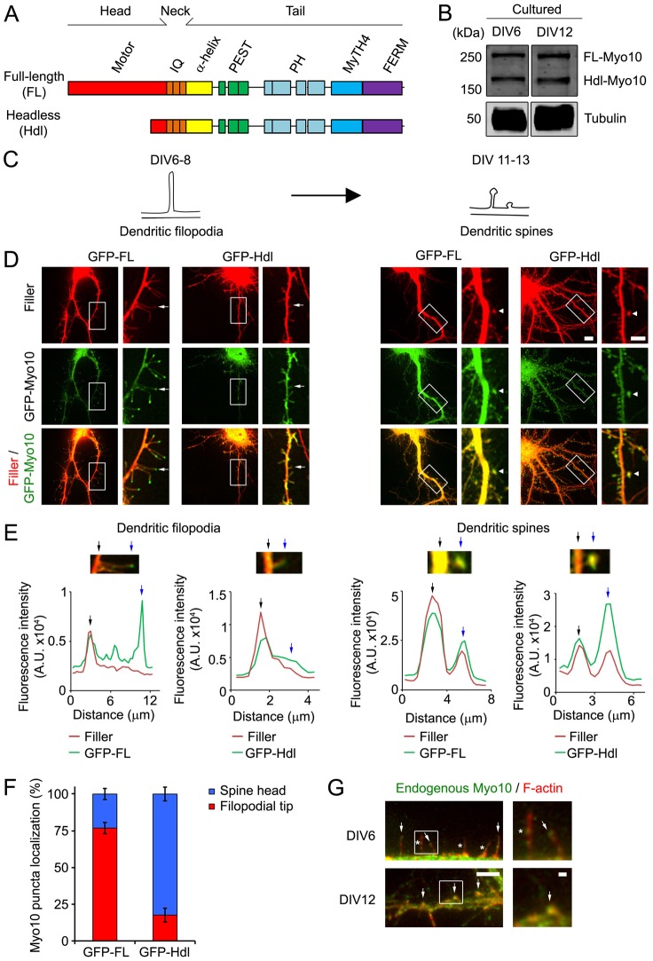
FL-Myo10 localizes to the tips of dendritic filopodia, whereas Hdl-Myo10 is enriched in spine heads. (A) A schematic illustrating the domains of Myo10 isoforms. (B) Cell lysates from DIV6 and DIV12 cultured hippocampal neurons, which were isolated from E19 rat embryos, were subjected to SDS-PAGE and immunoblotted for Myo10 (upper panels) and α-tubulin (lower panels) as a loading control. (C) A schematic denoting the time-period of spine development used in this study. Dendritic filopodia, which are precursors of dendritic spines, are the prevalent type of protrusions at DIV6–DIV8, whereas dendritic spines are predominant at DIV11–DIV13. Spines comprise a bulbous head that connects either directly to the dendritic shafts (on right) or by means of a thin neck (on left). (D) Neurons were co-transfected at DIV6 with either GFP–FL-Myo10 or GFP–Hdl-Myo10 and a fluorescent filler, mCerulean. Cells were then subjected to live-cell imaging one and six days after transfection to visualize dendritic filopodia and spines, respectively. mCerulean fluorescent filler is false-colored red. Scale bar: 10 µm. Higher magnification images of the boxed regions are shown on the right. Scale bar: 5 µm. Dendritic filopodia (arrows) and spines (arrow heads) are indicated. (E) To illustrate the localization of Myo10 isoforms to the tips of dendritic filopodia and to dendritic spines, linescan analyses were performed. The fluorescence intensities of the fluorescent filler (red) and GFP–Myo10 (green) through the dendrites and along the extended protrusions were graphed as a function of distance. The base (black arrow) and the tip (blue arrow) of dendritic protrusions are indicated. (F) Neurons (DIV5–DIV6) were transfected with GFP–FL-Myo10 and GFP–Hdl-Myo10, and the subcellular localization of the Myo10 isoforms was examined at DIV8. Quantification of the percentage of GFP–FL-Myo10 and GFP–Hdl-Myo10 puncta that localize to the tips of dendritic filopodia and spine heads is shown. Error bars represent the s.e.m. for 26 dendrites from three independent experiments. (G) DIV6 (top panels) and DIV12 (bottom panels) neurons were co-immunostained for endogenous Myo10 (green) and F-actin (red) to visualize dendritic filopodia and spines. Endogenous Myo10 localizes to the tips (arrows, top panels) and shafts (asterisk, top panels) of dendritic filopodia in DIV6 neurons. Myo10 is enriched in spine heads in DIV12 neurons (arrows, bottom panel). Scale bar: 5 µm. Higher magnification images of the boxed regions are shown (right panels). Scale bar: 1 µm.

