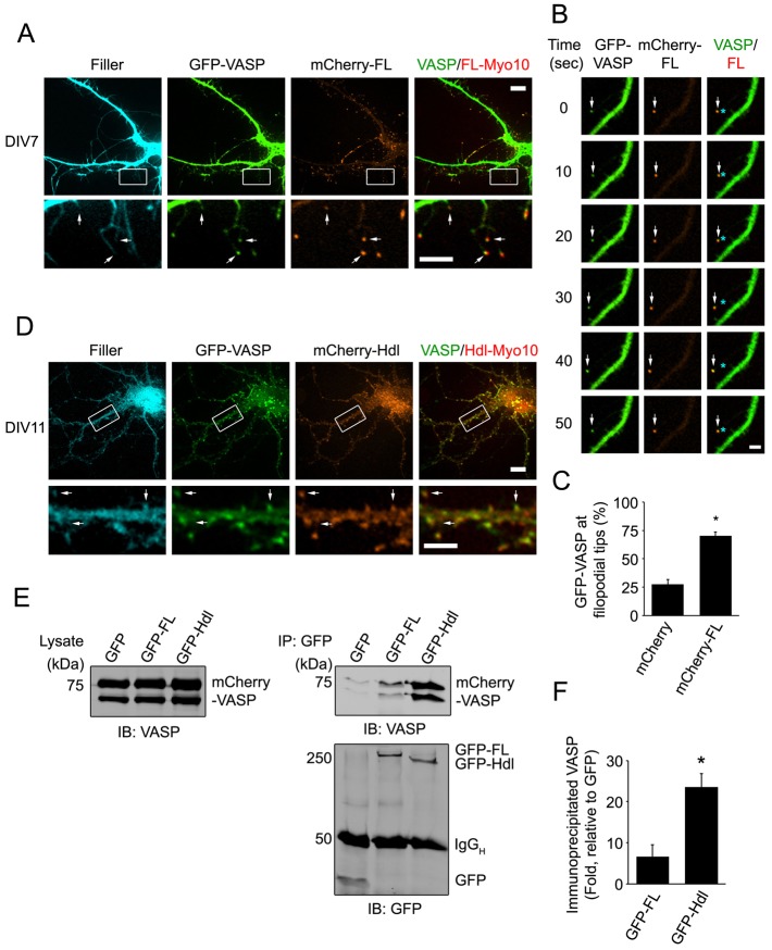
Fig. 6.
VASP colocalizes with both FL-Myo10 and Hdl-Myo10. (A) Neurons were co-transfected with GFP–VASP, mCherry–FL-Myo10 (mCherry-FL) and a fluorescent filler, mCerulean, at DIV6 and then subjected to live-cell imaging at DIV7 (upper panels). Scale bar: 10 µm. Higher-magnification images of the boxed regions are shown (lower panels). Scale bar: 5 µm. GFP–VASP and mCherry–FL-Myo10 puncta at the tips of dendritic filopodia are indicated (arrows). (B) A dendritic filopodium from a neuron transfected with GFP–VASP and mCherry–FL-Myo10 is shown. The mosaic of images shows a GFP–VASP punctum co-trafficking with mCherry–FL-Myo10 (arrows). Asterisks denote the original position of VASP or FL-Myo10 puncta. Scale bar: 1 µm. (C) Neurons were co-transfected with GFP–VASP, a fluorescent filler, mCerulean, and either mCherry or mCherry–FL-Myo10. A quantification of GFP–VASP localization (percentage) to dendritic filopodia tips from neurons expressing mCherry or mCherry–FL-Myo10 is shown. Error bars represent the s.e.m. for 30–36 cells from three separate experiments (*P <0.0001). (D) Neurons were co-transfected with GFP–VASP, mCherry–Hdl-Myo10 (mCherry-Hdl) and a fluorescent filler, mCerulean, at DIV6 and then subjected to live-cell imaging at DIV11 (top panels). Scale bar: 10 µm. Higher magnification images of the boxed regions are shown (lower panels). Scale bar: 5 µm. GFP–VASP and mCherry–Hdl-Myo10 puncta at spine heads are indicated (arrows). (E) HEK-293T cells were co-transfected with mCherry-VASP and either GFP, GFP–FL-Myo10 or GFP–Hdl-Myo10. 24 hours after transfection, cells were treated with cytochalasin D (2 µm) for 1 hour and then lysed. GFP and GFP–Myo10 isoforms were immunoprecipitated from cell lysates and subjected to immunoblot analysis. mCherry–VASP from total lysates (left panel) and immunoprecipitated complexes (upper right panel) were detected using an antibody against VASP. GFP and GFP–Myo10 isoforms from immunoprecipitated complexes were visualized with an antibody against GFP (lower right panel). (F) Quantifications of immunoprecipitated VASP from cells transfected with GFP–FL-Myo10 or GFP–Hdl-Myo10 are shown. Error bars represent the s.e.m. from three separate experiments (*P<0.02).

