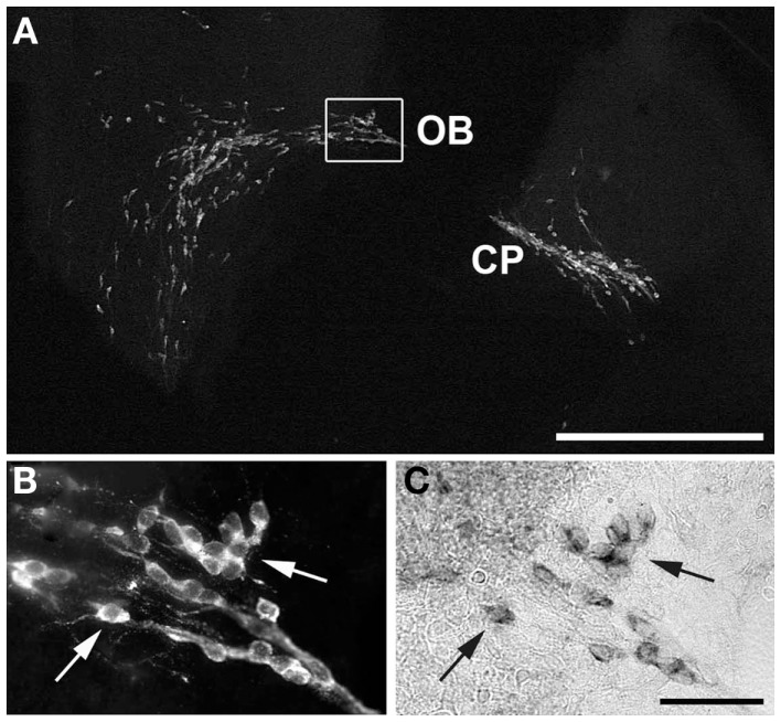
Figure 2.
Co-localization of GnRH and Kiss1r in migrating neurons. Kiss1r and GnRH were co-localized in migrating GnRH neurons in E15 mice. (A) shows a digital image of a fluorescent IHC for GnRH in a sagittal section from an E15 female also positive for Kiss1r. (B) (GnRH) and (C) (Kiss1r) show higher magnification images of the boxed area in (A), with arrows to indicate co-expressing cells. Compare the GnRH pattern in A to the Kiss1r pattern seen in the bottom right corner of Figure 1. CP, cribriform plate; OB, olfactory bulb. Scale Bars: white = 500 μm, black = 100 μm.

