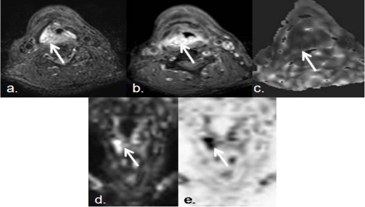
Figure 5.
Seventy two year old male, with squamous cell carcinoma of the right piriform sinus.Despite his small size, this malignant lesion associated marked diffusion restriction. (a,b) Pre- and postcontrast axial SPIR TSE T1-weighted images show a small enhancing and infiltrating lesion in right piriform sinus (white arrows); (c) Axial ADC map, (d) DWIBS coronal MIP, and (e) DWIBS inverted MIP (pseudo-PET) show high increased signal in DIBWS sequence and decreased signal in ADC map, that confirms diffusion restriction of the lesion (ADC: 0.8 × 10⁻³ mm2/s). DWI and ADC can be considered in routine clinical practice as complementary sequences to anatomical ones, in order to increase diagnostic accuracy.

