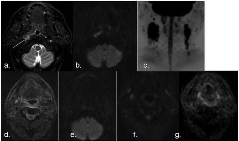
Figure 6.
Pre- and post-reatment SCC with lymph node metastasis. (a) Transverse STIR shows a small nodular hyperintense lesion in right torus tubarius; (b) The lesion demonstrates increased signal intensity on DWI with high b value (white arrow); (c) Coronal MIP of a DWIBS sequence with high b value demonstrates several bilateral enlarged lymph nodes with restricted diffusion The lesion was biopsied and confirmed as a SCC with bilateral metastatic lymph nodes. Postreatment follow-up MRI (six months after radio and chemotherapy); (d) axial STIR; (e and f) axial DWI with high b value at two different levels; (g) corresponding ADC map, demonstrates absence of recurrence, with no areas of restricted diffusion or suspicious lymph nodes.

