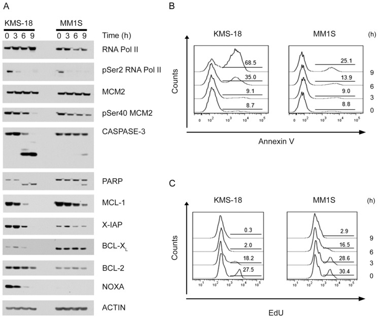
Figure 3.
Analysis of pro- and anti-apoptotic proteins in response to PHA-767491. KMS-18 and MM1S myeloma cells were incubated with 5 μM PHA-767491 for the indicated time. Protein extracts were prepared and analyzed by immunoblotting using the indicated antibodies (A). In parallel samples, apoptosis (B) and DNA synthesis (C) were analysed by flow cytometry using AV staining and EdU incorporation assay respectively. Numbers in the gated regions represent the percentage of cells positive for either AV (B) or EdU (C) staining.

