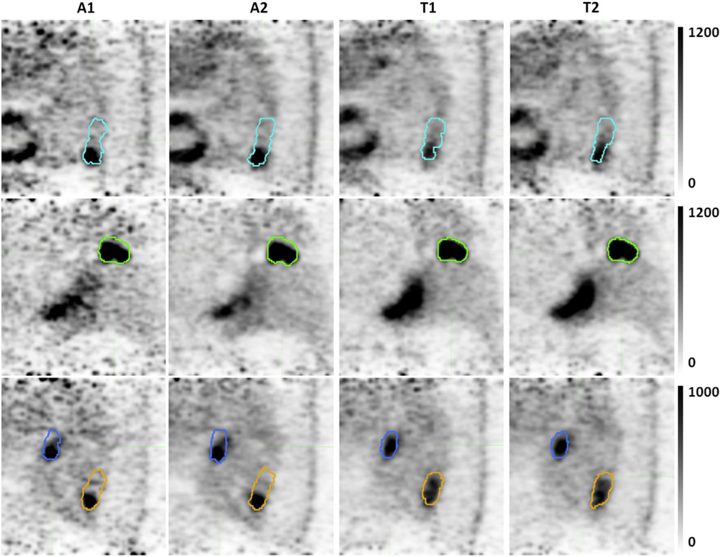
Fig. 4.
Selected slices highlighting algorithm differences in image generation and segmentation. Internal target volumes of tumor 3 (cyan), tumor 5 (green), lymph node 9 (blue), and tumor 9 (orange) registered to the end-inhale breathing phase. Pixel units are proportional to counts per second. Gated images from temporal-based algorithms have increased motion blurring artifacts.

