Figure 4.
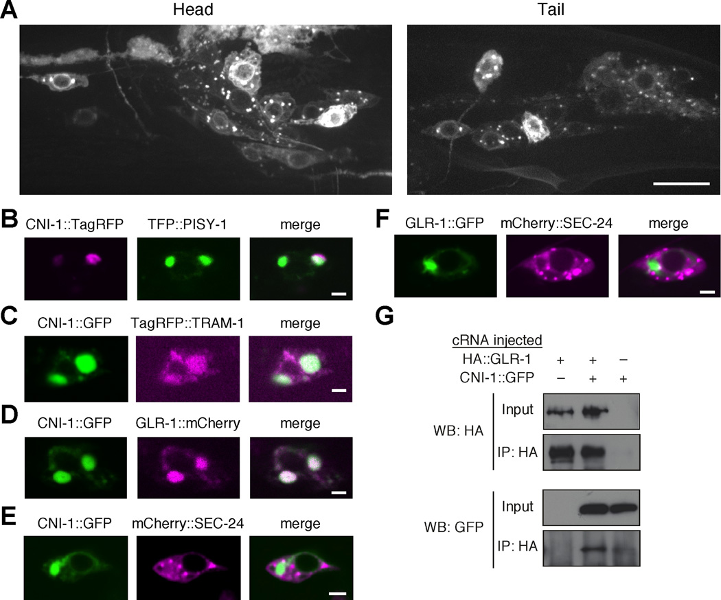
CNI-1 is widely expressed in the nervous system where it colocalizes with GLR-1 in the ER. (A) Confocal images of the head and tail region of transgenic worms that expressed the CNI-1 ::GFP reporter shown in Figure S5A . Scale bar represents 10 µm. (B–F) Single plane, confocal images of the AVA cell bodies in transgenic worms that expressed various combinations of fluorescently labeled proteins. Scale bars represent 2 µm. (G) Immunoprecipitation of HA::GLR-1 and CNI-1::GFP coexpressed in Xenopus oocytes.
See also Figure S5.

