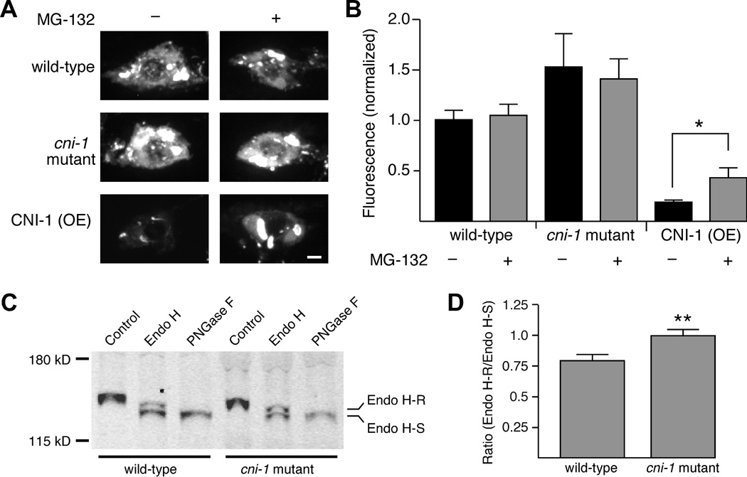
Figure 6.
Overexpression of CNI-1 results in GLR-1::GFP accumulation in neuronal cell bodies and its subsequent degradation. (A) Confocal images of GLR-1::GFP in AVA cell bodies in worms either with or without MG-132 treatment. Scale bar represents 2 µm. (B) Average GFP fluorescence intensity in worms either with (wild type, n=20; cni-1 mutant, n=12; CNI-1 (OE), n=15) or without (wild type, n=22; cni-1 mutant, n=13; CNI-1 (OE), n=16) MG-132 treatment. * p<0.05. (C) Western blot showing the relative amounts of Endo H sensitive (Endo H-S) and resistant (Endo H-R) GLR-1::GFP isolated from transgenic wild-type worms and cni-1 mutants. (D) The ratio of Endo H-R to Endo H-S GLR-1::GFP in wild type (n=11) and cni-1 mutants (n=10). ** p<0.01. Error bars represent SEM.

