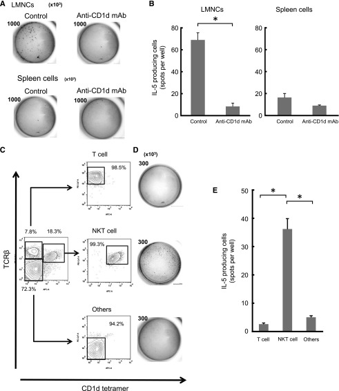
Figure 6.
NKT cells were predominant sources of IL-5 secreted after immunization with group A-RBCs. WT CD1d+/+ Balb/c mice received intraperitoneal injection of anti-mouse CD1d mAb (n = 3). Mice that received injections of isotype-matched Ab served as controls (n = 3). The mice were immunized with human blood group A-RBCs (5 × 10⁸/mouse) on day 1 after mAb administration. The mice were sacrificed to determine the IL-5–producing cells 6 hours after immunization. (A-B) LMNCs and spleen cells were seeded. The representative pictures of ELISPOT wells are shown (A) and the frequency of IL-5 producing cells is (B). Number in each picture refers to the total cells seeded per well (×103). (C-E) Six hours after immunization with A-RBCs, the LMNCs were isolated from CD1d+/+ Balb/c mice (n = 12). The pooled cells were used in ELISPOT assay to determine the frequency of IL-5–producing cells. The LMNCs were stained with APC-conjugated anti-mouse CD1d-tetramer and PE-Cy7–conjugated anti-mouse TCRβ. NKT cells (CD1d-tetramer+, TCRβ+), T cells (CD1d-tetramer–, TCRβ+), and the others (CD1d-tetramer–, TCRβ–) were isolated by sorting with FACS Aria. After sorting, the purities of NKT, T, and other cells were reanalyzed by FCM. (D-E) The representative pictures of ELISPOT wells are shown (D) and the frequency of IL-5–producing cells is shown (E). Number in each picture refers to the total cells seeded per well (×103). The results shown are the average ± SEM calculated from red spot number in quadruplicate wells. The results are representative of 2 similar experiments. *P < .05.

