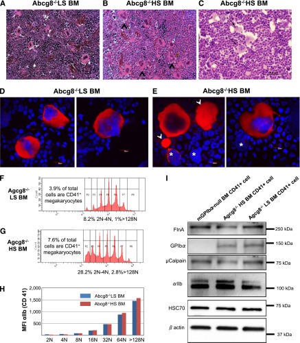
Figure 7.
Analyses of Abcg8−/− HS and Abcg8−/− LS megakaryocytes. (A-C) BM from Abcg8−/− LS (A) and Abcg8−/− HS mice (B-C) was harvested for histologic analysis. The number of multinucleated mature megakaryocytes (black arrowheads) was visually identified and counted in hematoxylin and eosin–stained sections. (C, black arrows) Emperipolesis of neutrophils into megakaryocytes. (D-E) BM megakaryocytes isolated by discontinuous gradient centrifugation over bovine serum albumin were stained with anti-αIIb mAb (red) and 4,6 diamidino-2-phenylindole (blue). Large αIIb-positive cytoplasmic extrusions (arrowhead) and “bare” megakaryocyte nuclei (asterisks) were noted in the BM samples from Abcg8−/− HS (E) but not in Abcg8−/− LS mice. (F-H) DNA ploidy profiles and integrin αIIb expression in BM megakaryocytes isolated using anti-CD41 magnetic beads and cultured in the presence of thrombopoietin for 60 hours. An HS diet resulted in a greater total number of CD41+ cells (7.6% vs 3.9%), with a corresponding proportional increase in both the high (>128N) and low (2N-4N) ploidy cells (F-G). Expression of integrin αIIb was similar in Abcg8−/− HS and Abcg8−/− LS cells regardless of ploidy (H). (I) Western blot analysis of purified, CD41-positive megakaryocytes. Expression of filamin A (FlnA), GPIbα, and µ-calpain in megakaryocytes derived from Abcg8−/− HS mice was slightly decreased compared with that of Abcg8−/− LS mice. No significant differences were observed in the expression of HSC70 and β actin.

