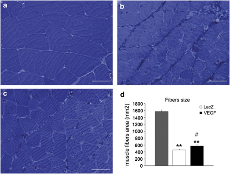
Figure 4.
Light microscopy of toluidine blue-stained semithin sections of a normal muscle (a) and denervated muscle 1 month after surgery and treatment with AAV-LacZ (b) or AAV-VEGF (c). Histogram in d shows the results of morphometrical evaluation of muscle fibers size (number of rats analyzed=17). #P⩽0.05 VEGF vs LacZ; **P⩽0.01 VEGF and LacZ vs control (CTRL). Scale bars: 25 μm.

