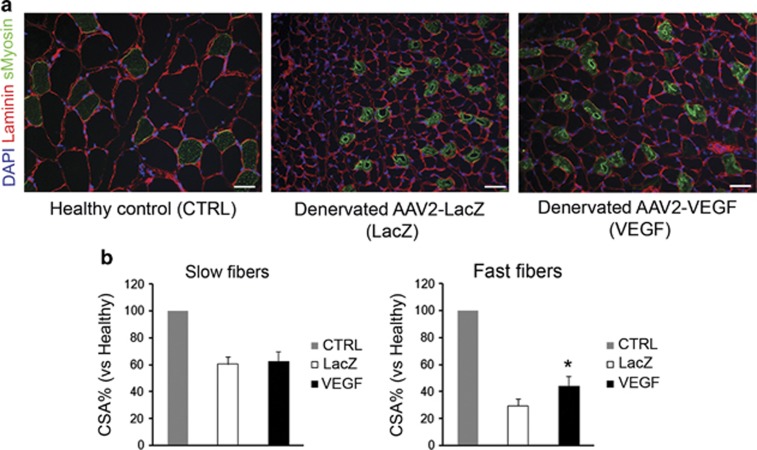
Figure 5.
Muscle sections were stained in immunohistochemistry with laminin (red) and slow myosin (green) in order to study the effect of VEGF overexpression on skeletal muscle fiber cross-sectional area. Cellular nuclei were counterstained with 4',6-diamidino-2-phenylindole (DAPI; blue). Scale bars: 25 μm (a). Histograms that represent quantification of the fiber cross-sectional area (b); results are expressed as a percentage compared with the matched fiber type in the contralateral healthy muscle (number of rats analyzed=6). *P⩽0.05 VEGF vs LacZ.

