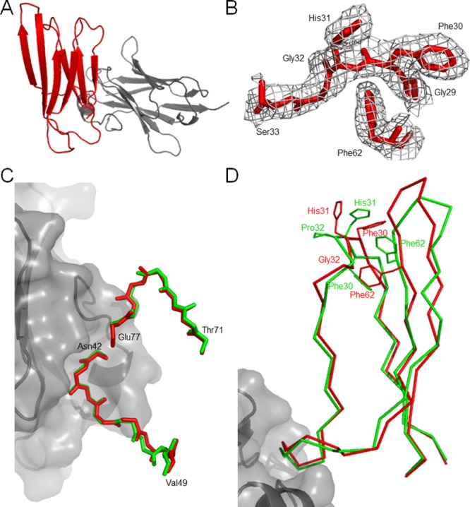
Figure 3.

Crystal structure of P32G β2m•Nb24 complex. (A) The P32G β2m•Nb24 complex as a stable heterodimer with P32G β2m in red and Nb24 in gray. (B) The 2Fo – Fc omit density map is shown for residues Gly29, Phe30, His31, Ser33, and Phe62. The map is contoured at 1 σ within 1.6 Å of the residues. (C) Comparison between the P32G β2m•Nb24 structure (red) and the β2m structure in the MHC I complex (PDB entry 1DUZ25;green) focusing on the high similarity of the main-chain conformation in the binding region of P32G β2m to Nb24 (in gray surface representation). (D) Comparison between P32G β2m•Nb24 structure (red) and the β2m structure (PDB entry 1DUZ25; green) focusing on the key structural consequences of the cis–trans isomerization at position 32.
