
Fig. 1.
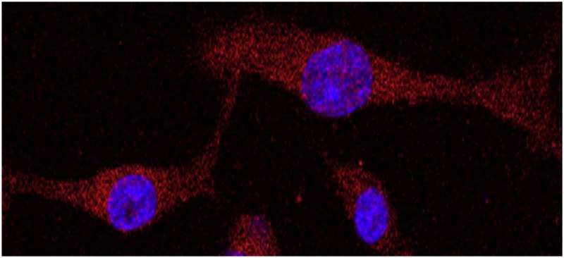
Representative immunocytochemical image of isolated primary cell lines demonstrating that these cultures are glioma cells. Nuclei are stained with 4,6′-diamidino-2-phenylindole (blue) and cells with glial fibrillary acidic protein (GFAP) (red) and imaged using confocal microscopy at (×40) magnification. The large nuclear to cytoplasmic ratio suggests that these cells are neoplastic, and the lack of GFAP-negative cells suggests that these are not neurons in culture. (This figure is available in colour at www.sciencedirect.com)