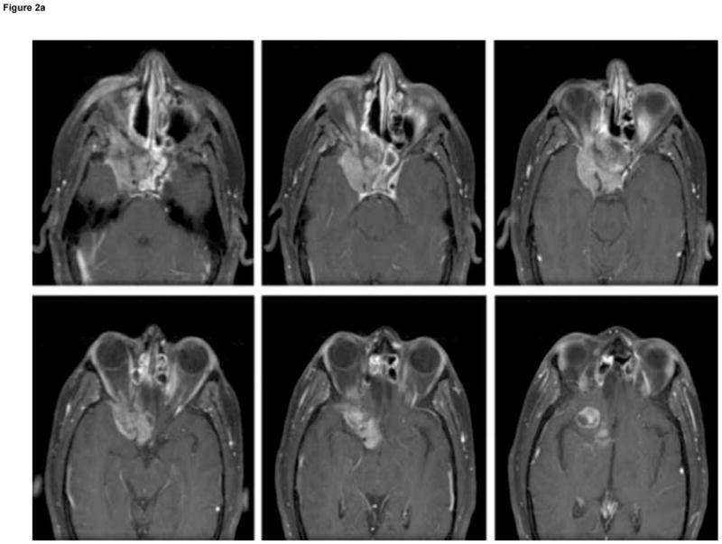
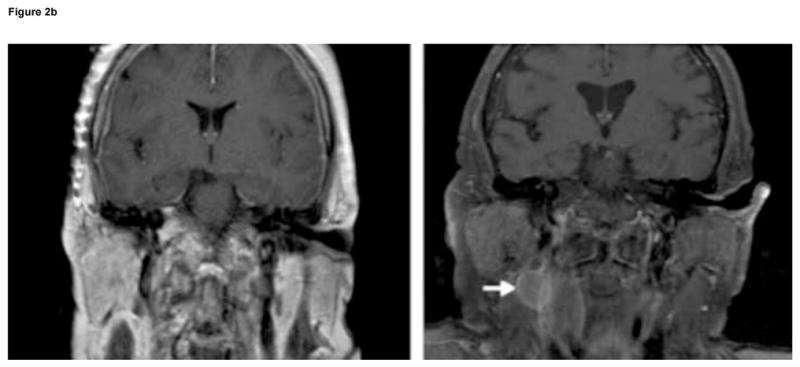
Figure 2.


Patterns of recurrence in two patients with high-grade
esthesioneuroblastoma (EN): (A) six sequential axial, T1-weighted, post-gadolinium MRI of Patient 6 with high-grade, aggressive intranasal and intracranial recurrence involving the cavernous sinus and frontal lobe, 41 months after primary resection of a high-grade EN; and (B) coronal, T1-weighted, contrast-enhanced MRI of Patient 14 showing absence of nodal metastasis before initial surgery (left), and evidence of nodal metastasis (white arrow) 48 months after first surgery (right).
