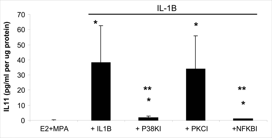
Figure 5. Involvement of NFκB, p38 MAPK and PKC signaling in IL-1β-enhanced IL-11 output by decidual cells maintained in E2 + MPA.
Confluent, leukocyte-free first trimester decidual cells were incubated for 7 days in 10−8 M E2 + 10−7 M MPA, then switched to DM with the steroids alone ± 1ng/ml IL-1β with and without an NFκB inhibitor (Activation Inhibitor III, NFκBI) at 10−5M, or a p38 MAP kinase inhibitor (SB203580; p38KI) at 10−5M, or a PKC inhibitor (Calphostin C, PKCI) at 10−7M. Bars represent mean ± SEM IL-11 levels as pg/ml/µg cell protein as measured by ELISA in conditioned DM and normalized to cell protein. (n=4 separate patients’ decidual specimens). * versus E2 + MPA; p<0.05.

