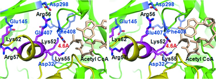
Figure 4.

Enlarged stereoview of the active binding site in the docking model. Side-chains (in blue) and acetyl-CoA (in wheat color) of Mtb Eis (backbone in green and residues labeled in blue). Side-chains (in magenta) of DUSP16/MKP-7 (backbone in yellow and residues labeled in black) are shown as a ball-and-stick model.
