
Figure 2. TBBPA effect on DCF fluorescence in HBSS solution.
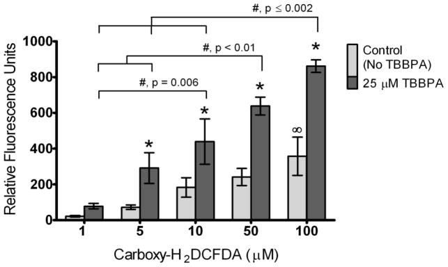
DCF fluorescence was assessed in cell-free and serum-free HBSS solutions containing 1, 5, 10, 50 or 100 μM DCFH2DA in either the absence (controls) or presence of 25 μM TBBPA. Controls were incubated with solvent only (0.1% DMSO). Columns represent means ± SE of 3 independent experiments containing 6 replicates each. There was a significant TBBPA * Carboxy-DCFH2DA interaction (ANOVA, p=0.03). *, Statistically significant increase compared to controls within a particular concentration of carboxy- H2DCFDA (p<0.05). ∞, Statistically significant increase in controls at 100 μM carboxy-H2DCFDA compared to controls at 1 μM carboxy-H2DCFDA. #, Statistically significant differences between samples incubated with TBBPA at different concentrations of carboxy-H2DCFDA (p-values shown on graph).