
Figure 1. Construction and detection of the 3xSTaA14Q-tmLT toxoid fusion.
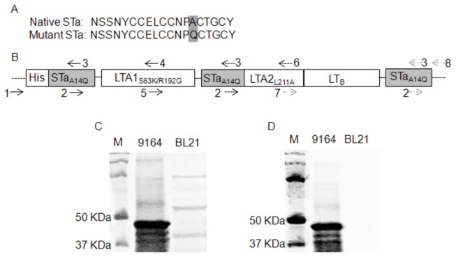
Panel A: Amino acid sequences of the native STa toxin and the STaA14Q toxoid. Panel B: Construction of the 3xSTaA14Q-tmLT toxoid fusion gene with 3 copies of the STaA14Q toxoid gene genetically fused at the 5’ end, within LTA, and the 3’ end of the tmLT toxoid gene (LTS63K/R192G/L211A). The numbers and arrows indicated primers used in PCRs to mutate the genes and to amplify fragments to be overlapped for a single open reading frame encoding the 3xSTaA14Q-tmLT toxoid fusion. This chimeric toxoid fusion gene was cloned in vector pET28α and expressed in E. coli BL21 as a 6xHis-tagged fusion protein. The drawing scale is not in proportion to nucleotide fragment sizes. Panel C: Detection of 6xHis-tagged 3xSTaA14Q-tmLT toxoid fusion protein in Western blot using 12% PAGE gel, with rabbit anti-CT antiserum (1:3300; Sigma) and IRDye-labeled goat anti-rabbit IgG (1:5000; LI-COR, Lincoln, NE). Panel D: Detection of the toxoid fusion protein with purified rabbit anti-STa antiserum (1:5000) and IRDye-labeled goat anti-rabbit IgG (1:5000; LI-COR, Lincoln, NE). Extracted 6xHis-tagged protein form fusion strain 9164 and total protein extracts from negative control strain 8955 were examined in the SDS-PAGE. Lane M is the protein marker (Precision Plus Protein Pre-stained standards, Bio-Rad, Hercules, CA).