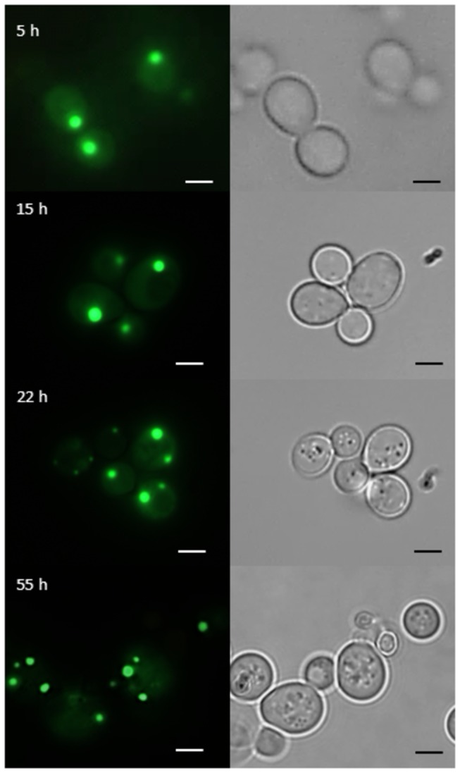
Figure 5. Subcellular localization of Gph1p-GFP by fluorescence microscopy.

Fluorescence microscopy was performed as described in the legend to Figure 2. GFP fluorescence (left panel) and transmission microscopy (right panel) of cells bearing a Gph1p-GFP hybrid are shown. Cells were grown in minimal medium –ura –met at 30°C and pictures were taken from cells at early (5 h), middle (15 h) and late (22 h) exponentially phase and at the late stationary phase (55 h). The size of the scale bar is 1 µm.
