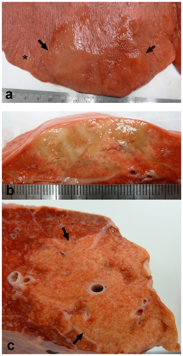
Figure 1. Experimental EHV 5 infection; gross pathology (horse E3, E4).

Nodules of fibrosis (A, between arrows, and at asterisk; horse E4) were evident beneath the pleura (cut section, B; horse E4). The nodules of fibrosis extended into the underlying alveolar parenchyma (B,C; horse E3,E4 respectively).
