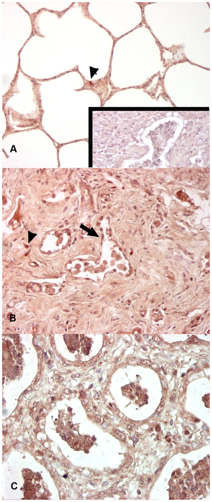
Figure 5. Immunohistochemistry for EHV 5 (horse C2, E4).

Control horse (C2) has detectable EHV 5 antigen within scattered alveolar macrophages (arrowhead). EHV 5 infected horses (B) had detectable EHV 5 antigen within the nodules of fibrosis, including the honeycomb epithelial cells (arrow), interstitial fibroblasts, and macrophages. Similar distribution of EHV 5 was found in the lungs of naturally-acquired EMPF (C). Inset (A) negative control, rabbit serum IgG substituted for primary antibody. Magnification: A-C - 20x.
