
Figure 2. Structure and nuclear reorganization of BCL-6/BCoR inclusions.
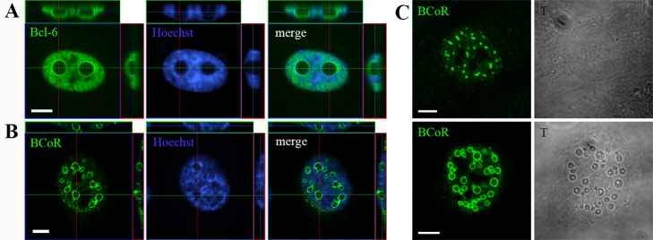
ECs were transfected with EFp-BCL-6 (A) or EFp-BCoR-A (B and C) expression plasmid and processed with Hoechst 33342 DNA stain and antibodies against BCL-6 or BCoR for CLSM imaging after 24 hours. (A and B) Compilation of Z-stack series into orthogonal projections. (C) Comparison between fluorescence and light transmission (gray scale) images of BCoR-A transfected cells. Scale bars: 5 µm.