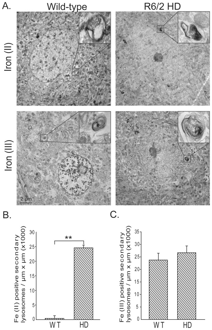
Figure 2. Iron (II) accumulates in R6/2 HD striatal neurons.
Iron (II) and iron (III) were determined by a modification of the perfusion Turnbull’s and Perl’s iron stains (see methods). A. Electron photomicrographs show striatal neurons with foci of iron (II) or (III) staining in membrane-bound structures consistent with secondary lysosomes. Quantification of iron (II) (B) and iron (III) (C) staining reveals significantly elevated iron (II) while iron (III) is unaltered. P-value: **< 0.01, n=2 and 5 neurons / mouse .

