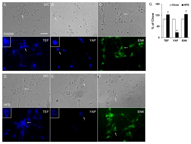
Figure 4. Isolated hypothalamic neurons from E19 embryos.
Single-labeling immunocharacterization of dissociated hypothalamic neurons from chow embryos expressing (A) TEF, (B) YAP, or (C) ENK; and from HFD embryos expressing (D) TEF, (E) YAP, or (F) ENK. (G). In the HFD compared to chow embryos, there was a statistically significant decrease in the number of YAP-labeled neurons and an increase in the number of TEF- and ENK-labeled neurons. Top set of images is neurons from chow embryos, bottom set is neurons from HFD embryos. Arrows point to the same cell in the top and bottom image (DIC and fluorescence). Insert shows larger image of neuron with either cytoplasmic or nuclear localization of TEF or YAP. Results are expressed as means ± SEM (n = 4). *p < 0.05, compared with chow. ENK = enkephalin; scale bar is 25 µM.

