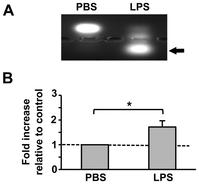
Figure 5. LPS stimulation induces PKC activity in mosquito cells.
Cell lysates from immortalized A. stephensi ASE cells stimulated with PBS as a control or with 100 µg/ml LPS for 30 min were incubated with fluorescently tagged C1 peptides (a PKC substrate) for 30 min and analyzed by agarose gel electrophoresis. Phosphorylated C1 peptides, which are directly correlated with PKC enzymatic activity, are indicated by the arrow. (A) Representative agarose gel image. (B) Means ± SEMs of phosphorylated C1 peptide fluorescence normalized to PBS treated controls, n = 4. Pairwise comparisons of treatments and matched controls were analyzed by Student’s t-test, *p<0.05.

