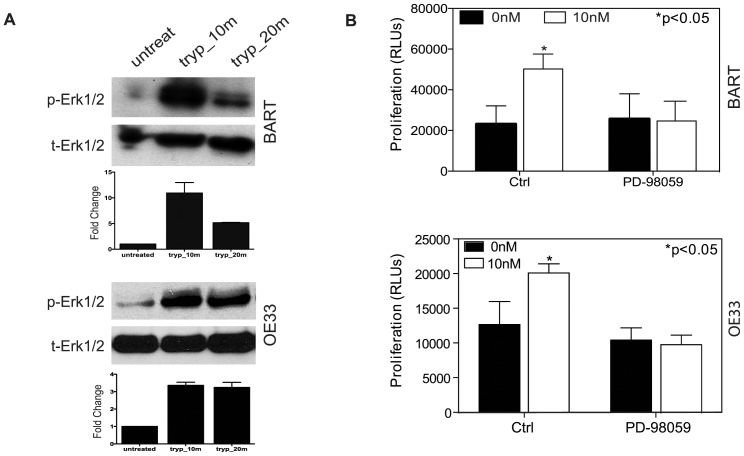
Figure 3. Erk/MAPK involvement in Trypsin Induced Signaling.
(A) Showing representative photographs for Western blot analysis of Erk1/2 phosphorylation status in BART and OE33 cells in the presence of or the absence of 10 nM trypsin at different time course as indicated. (B) Proliferation assay revealed that MEK inhibition abolished the proliferative responses of both BART and OE33 cells to 10 nM trypsin stimulation.

