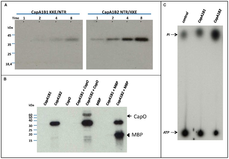
Figure 6. CapA1B1 activity assays. A.
– Effect of CapB2 N3T4R5 substitution for CapB1 K3K4E5 and vice versa on CaB1 and CapB2 kinase activity. CapA1B1(KKE/NTR) (left panel) and CapA1B2(NTR/KKE) (right panel) were incubated in the presence of radioactive ATP for either 1, 2, 4 or 8 minutes. After SDS-PAGE analysis, their autophosphorylation was visualized by autoradiography. B – CapO and myelin-binding protein (MBP) phosphorylation assays. The ability of CapA1B1 and CapA1B2 to phosphorylate CapO or the myelin-binding protein (MBP) was determined after incubation in the presence of radioactive ATP, SDS-PAGE analysis and film exposure. The arrow points to CapO whereas the arrowhead shows the MBP. C – ATPase activity of CapA1B1 and CapA1B2. The hydrolysis of [γ-32P] ATP was measured in the presence of either CapA1B1 or CapA1B2 as described under “Materials and Methods”. Migration of a control reaction mixture devoid of CapA1B1 or CapA1B2 is also shown.

