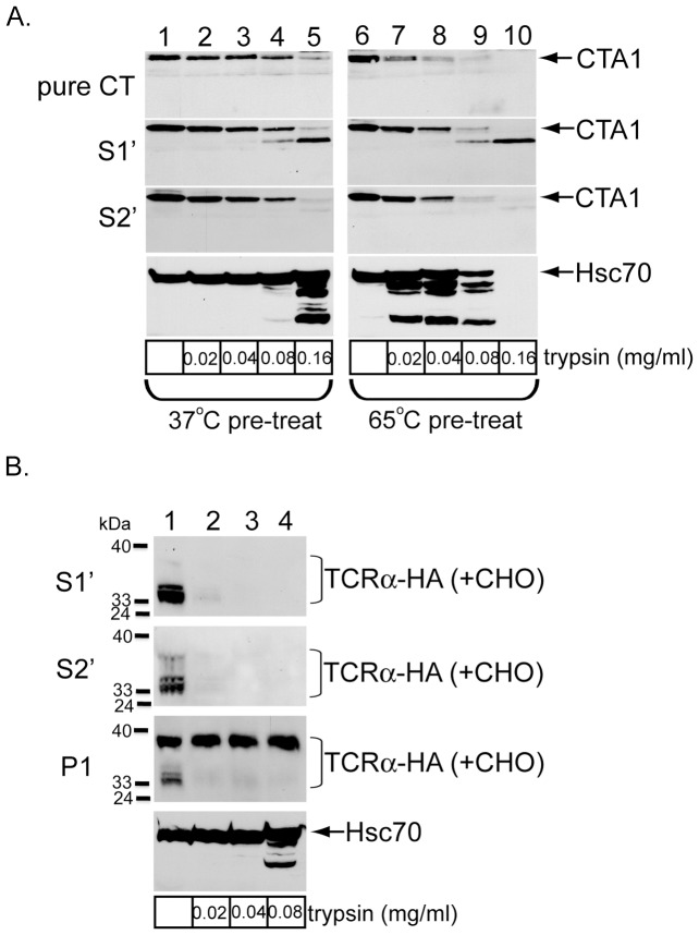
Figure 4. CTA1 but not TCRα released into the cytosol is largely folded.
(A) Purified CTA was incubated with CE to mimic the protein concentrations in S1′ and S2′, as well as 20 mM DTT to generate CTA1. S1′ and S2′ containing CTA1 were generated as before. Pure CTA1, S1′, and S2′ were incubated at 37°C (left panels) or 65°C for 15 minutes and then incubated with the indicated trypsin concentration for one hour at 4°C. Reactions were stopped with TLCK and sample buffer and the samples analyzed by immunoblotting with the indicated antibodies. (B) S1′, S2′, and intact P1 were generated from epoxomicin-treated cells expressing TCRα-HA. Samples were incubated with the indicated trypsin concentration for one hour at 4°C. Reactions were stopped and P1 lysed with sample buffer, and the samples analyzed by immunoblotting with the indicated antibodies.

