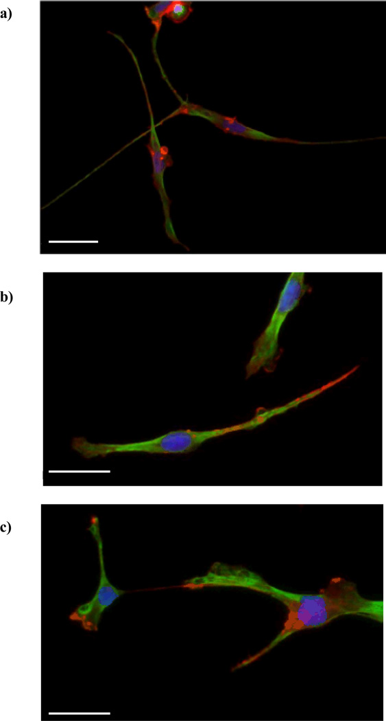
Figure 6.
Immunofluorescent images of PC12 cells following release from the hydrogel scaffolds. The cells were embedded in PEG hydrogels containing RGDS (a), YIGSR (b), or IKVAV (c) ligands (100 µM) and were released upon hydrogel degradation. The cells adhering to the collagen-coated coverslips (placed underneath the hydrogels) were fixed and stained 48 h after hydrogel degradation. Neurons were co-labeled for actin (red) and tubulin (green). Cell nuclei were labeled with DAPI (blue). Scale bars are 50 µm.

