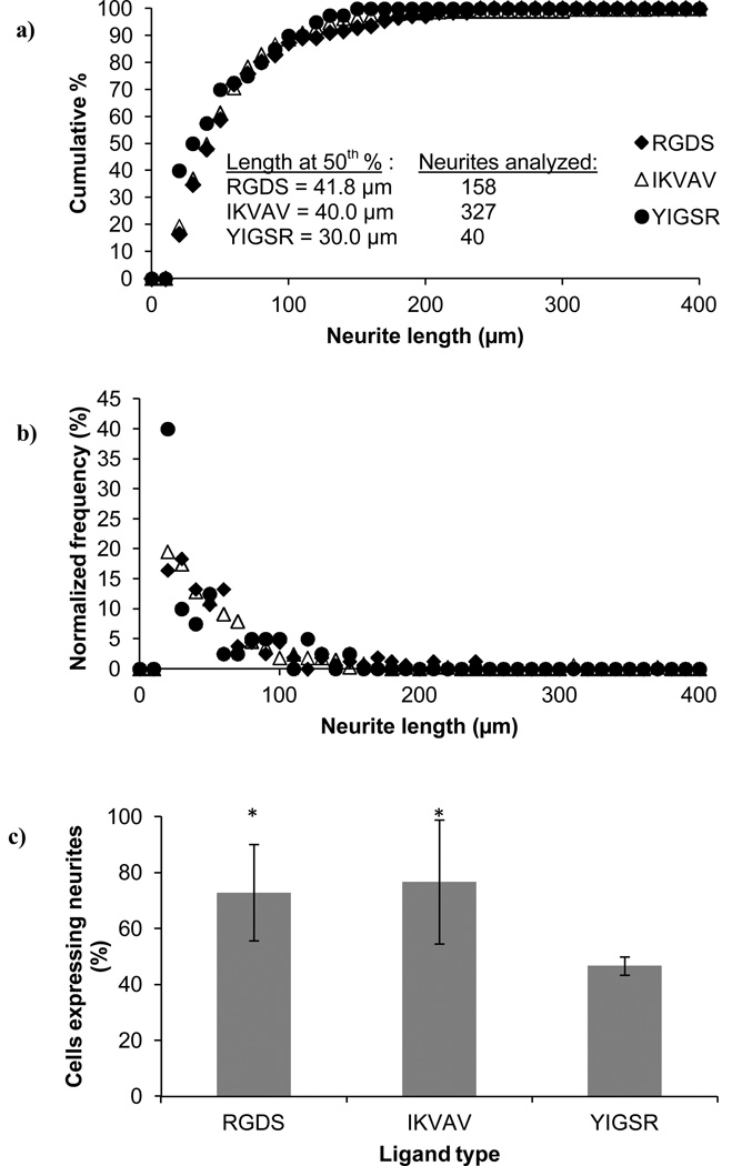
Figure 7.
Neurite extension following release from the PEG scaffolds. The cells were embedded in PEG hydrogels containing RGDS, IKVAV or YIGSR ligands (100 µM) and were released upon hydrogel degradation. Cell analysis were performed 48 h after the cells had been released from the hydrogels and adhered onto collagen-coated coverslips. Neurite length, represented by (a) the cumulative distribution and (b) the normalized frequency, as well as (c) the number of cells with neurites, revealed differences between the PC12 cells released from the YIGSR-modified PEG hydrogels, compared to the RGDS- or IKVAV-modified PEG hydrogels. Asterisks designate significant differences from YIGSR in (c).

