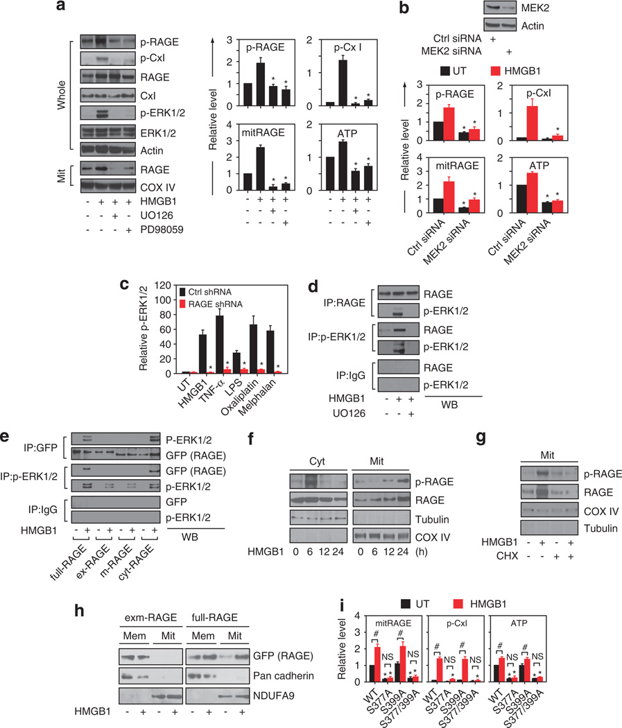
Figure 4.
Ser377 is required for RAGE activity in mitochondria. (a) Panc02 cells were treated with 10 µg/ml HMGB1 for 24 h in the absence or the presence of the ERK inhibitors U0126 and PD98059 (20 µm), followed by western blot analysis of whole cells or mitochondrial extracts. Quantitative data is shown in the right panel, with normal ctrl set as 1 or 0.1 (mean±s.d., n = 3, *P <0.001 versus single HMGB1 group). (b) Knockdown of MEK2 by siRNA in Panc02 cells decreased relative levels of p-RAGE, p-CxI, mitRAGE, and ATP with or without 10 µg/ml HMGB1 treatment for 24 h. Data is shown as mean±s.d., with the normal control set as 1 or 0.1 (n = 3, *P <0.001 versus ctrl siRNA group). (c) uantitative analysis of p-ERK in wild-type and RAGE knockdown Panc02 cells following treatment with a representative damage-associated molecular pattern molecule (HMGB1), pathogen-associated molecular pattern molecule (lipopolysaccharide), cytokine (TNFα) or chemotherapeutic drug (melphalan, oxaliplatin) as indicated for 24h (mean±s.d., n=3, *P <0.001 versus ctrl shRNA group). (d) Antibodies to RAGE, p-ERK1/2 or a nonspecific control IgG were incubated with Panc02 extracts following 10 µg/ml HMGB1 treatment with or without U0126 (20 µm) for 12 h. Immunoprecipitates were resolved on SDS–PAGE and probed for RAGE, p-ERK1/2. (e) Panc02 cells were transfected with full-RAGE, ex-RAGE, m-RAGE and cyt-RAGE plasmids fused with a GFP-tag. Antibodies to GFP, p-ERK1/2 or a nonspecific control IgG were incubated with Panc02 extracts as indicated. Immunoprecipitates were resolved on SDS–PAGE and probed for GFP, p-ERK1/2. (f) Panc02 cells were treated with 10 µg/ml HMGB1 for 24 h, followed by western blot analysis of cytosolic (‘Cyt’) or mitochondrial (‘Mit’) extracts as indicated. (G) Panc02 cells 10 µg/ml HMGB1 for 24 h in the absence or the presence of cycloheximide (CHX, 10 µg/ml), followed by western blot analysis of mitochondrial extracts. (h) Panc02 cells were transfected with full-RAGE and exm-RAGE plasmids fused with a GFP-tag, then treated with 10 µg/ml HMGB1 for 24 h. western blot analysis of membrane (‘Mem’) and mitochondrial (‘Mit’) extracts. (i) Panc02 cells were transfected with full-RAGE (‘WT’) and mutants as indicated, and treated with 10 µg/ml HMGB1 for 24 h. Relative levels of mitRAGE, p-CxI, and ATP are shown as mean±s.d., with normal ctrl set as 1 or 0.1 (n =3, *P <0.001 versus normal WT group, #P <0.01, NS: not significant).

