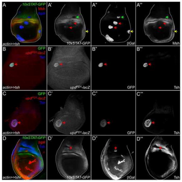
Figure 6. Stat92E and Tsh do not exhibit mutual antagonism.
(A) 10xSTAT-GFP is repressed in Tsh-clones residing in the proximal hinge (green arrow, A′, A″) but not in the lateral hinge (A′, A″, yellow arrow), despite Msh being repressed in both clones (A‴, green and yellow arrow). By contrast, 10xSTAT-GFP is induced in Tsh-expressing clones residing in the pouch (arrows in A′, A‴). Msh (red) is weakly induced in some Tsh-expressing clones in the pouch (red arrows in A‴). A′ is single channel for 10xSTAT-GFP; A″ for βGal; A‴ for Msh.
(B, C) An upd enhancer trap (updPD1, marked by βGal (red)) is cell-autonomously induced in Tsh-expressing clones (arrows in B–B‴ and C–C‴), which express both GFP (green) and Tsh (blue). The upregulation of upd occurs in both the hinge (B) and the pouch (B). Note that the endogenous domain of Tsh is not visible in B‴ because the gain of the confocal was decreased to not over-saturate the signal. B′ and C′ are single channel for βGal; B″ and C″ for GFP; B‴ and C″ for Tsh.
(D) Endogenous 10xSTAT-GFP is not altered in a Tsh RNAi (tshi)-expressing clone (marked by βGal (red)) residing in the proximal hinge (arrow in D′-D‴). The lack of Tsh protein within the clone (arrow in D‴) indicates the effective of the knockdown. Tsh is blue in D. D′ is single channel for 10xSTAT-GFP ; D′ for βGal; D‴ for Tsh.

