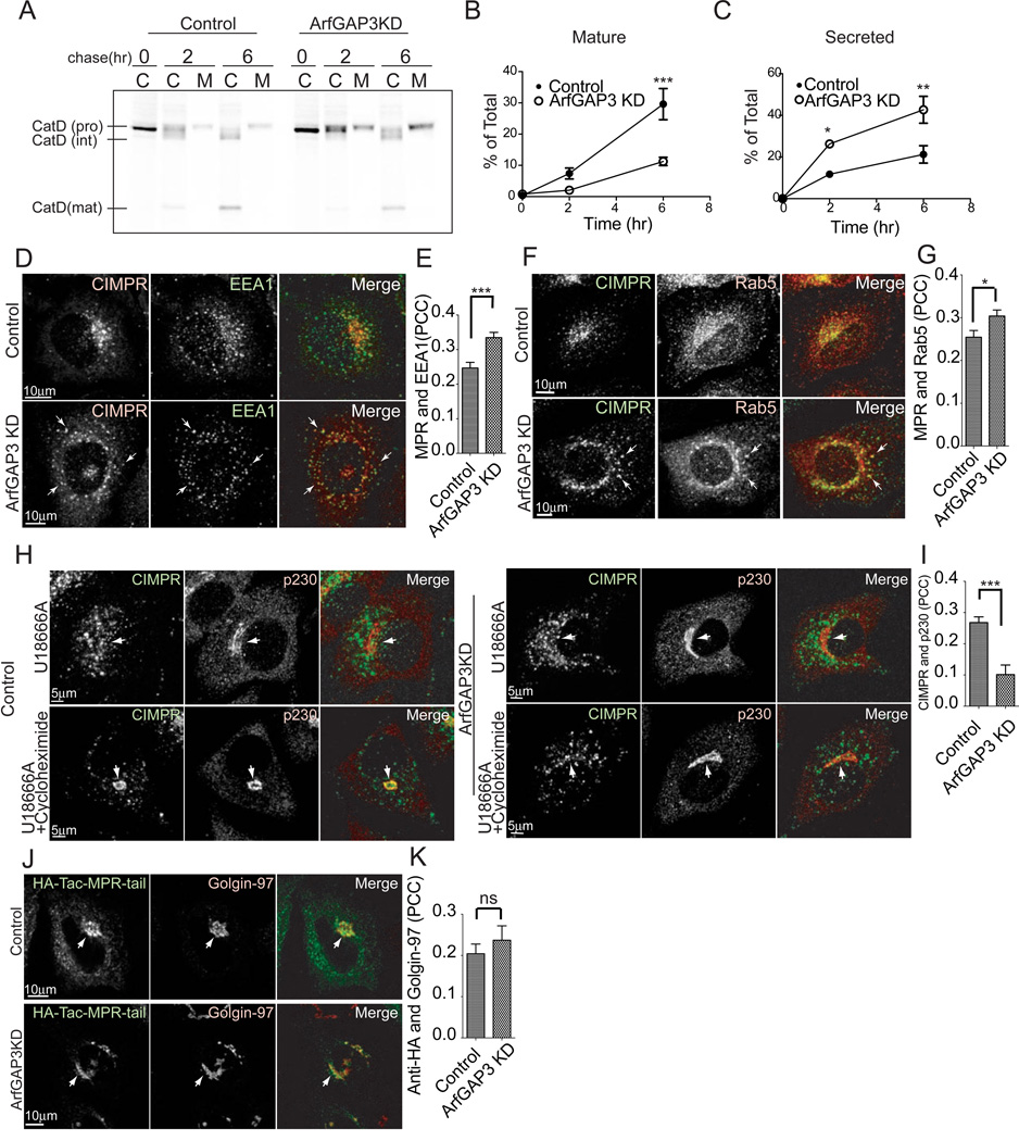
Figure 2. CIMPR transport is perturbed in ArfGAP3 KD cells.
A. Cells were pulse-labeled with [35S] methionine/cysteine for 1 hr, chased for 2 or 6 hr, and then cell-associated and medium-associated proteins were immunoprecipitated with anti-Cathepsin D antibody. A representative experiment out of three is shown. The lanes indicated C are the cell-associated and lanes labeled M are Medium-associated radiolabeled Cathepsin D. B. Mature Cathepsin D was decreased in ArfGAP3 KD cells. The amount of mature Cathepsin D detected in the autoradiograms presented in panel (A) was quantified. C. Quantification of pro-Cathepsin D detected in the cell culture media. More Cathepsin D was secreted in ArfGAP3 KD cells. D. Control and ArfGAP3 KD cells were double-stained with CIMPR and EEA1 antibodies. Arrows indicate colocalization of CIMPR with EEA1. E. Quantification of colocalization between CIMPR and EEA1. The average PCCs (n=30) are presented with S.E.M. F. Control and ArfGAP3 KD cells were double-stained with CIMPR and Rab5 antibodies. Arrows indicate colocalization of CIMPR with Rab5. G. Quantification of colocalization between CIMPR and Rab5. The average PCCs (n=30) are presented with S.E.M. H. Endosome to TGN transport of endogenous CIMPR is slowed in ArfGAP3 knockdown cells. Cells were treated with 3 µg/ml U18666A for 36 hr, thereby trapping CIMPR in endosomes. Cells were subsequently treated with 40 µg/ml cycloheximide for 3 hr, which results in the transport of CIMPR to the TGN in control cells but not in ArfGAP3 KD cells. The cells were stained with CIMPR and p230. Arrows indicate the Golgi area stained with p230. I. Quantification of colocalization between endogenous CIMPR and p230 in cells treated with U18666A and cycloheximide. The average PCCs (n=30) are presented with S.E.M. J. ArfGAP3 does not affect CIMPR lacking luminal domain. HeLa cells overexpressing HA-Tac-MPR-tail were incubated with anti-HA antibody for 3 min, washed and chased for 30 min. The cells were stained with Golgin-97. Arrows indicate HA-Tac-MPR-Tail localized at the Golgi. K. Quantification of colocalization between internalized anti-HA antibody and Golgin-97. The average PCCs (n=30) are presented with S.E.M. * indicates significant difference, p < 0.05; **, p < 0.01; ***, p < 0.001; ns, not significant.

