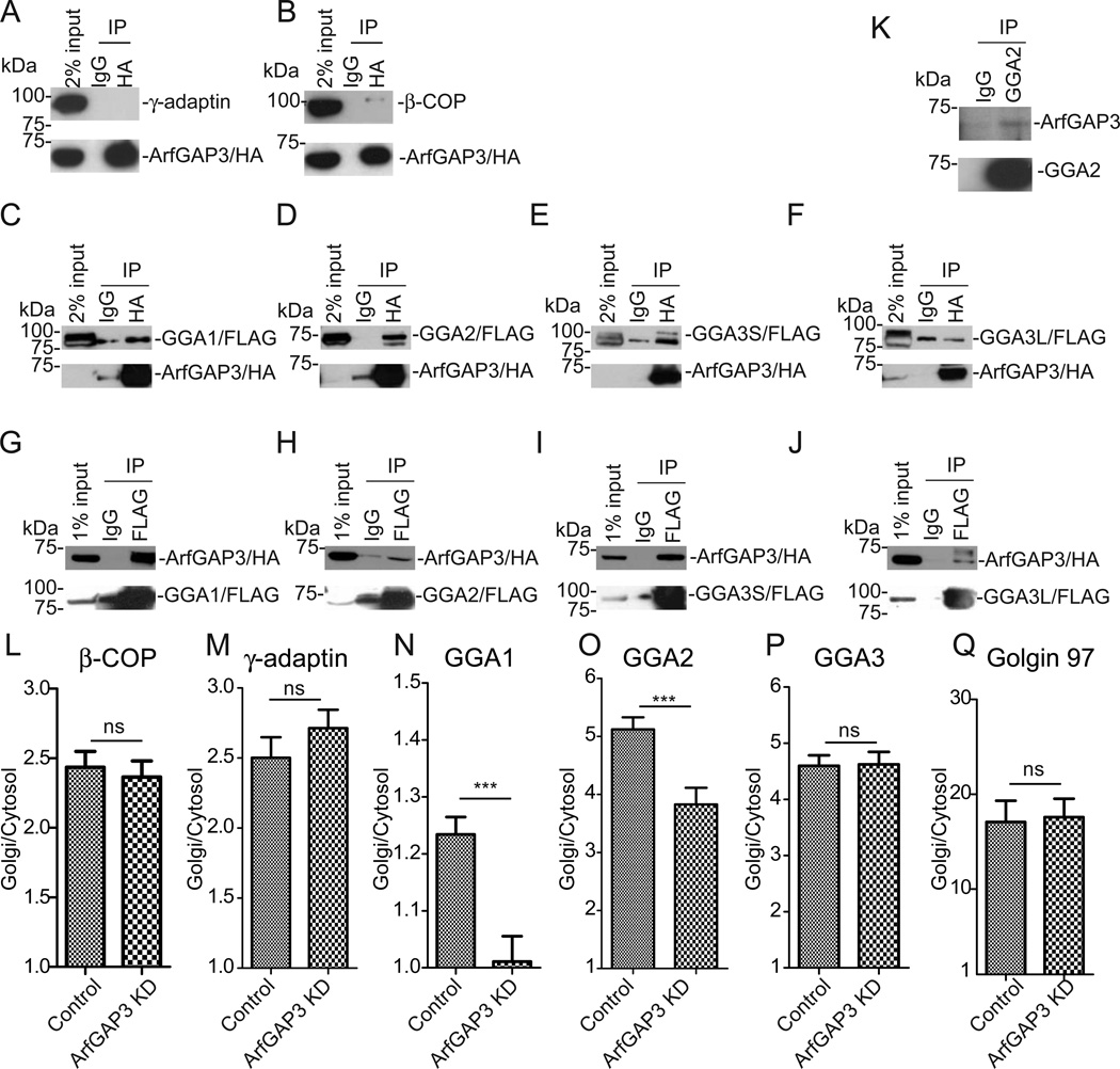
Figure 4. ArfGAP3 associates with GGAs.
A,B. 293T cells were transfected with HA-tagged ArfGAP3 and immunoprecipitated with anti-HA, blotted with anti-γ-adaptin (AP-1 subunit) (A) or anti-β-COP (COPI subunit) (B). C-F. 293T cells were double-transfected with HA-tagged ArfGAP3 and FLAG-tagged GGA1 (C), GGA2 (D), GGA3S (E), GGA3L (F), immunoprecipitated with anti-HA and blotted with anti-FLAG antibody. G-H. 293T cells were double-transfected with HA-tagged ArfGAP3 and FLAG-tagged GGA1 (G), GGA2 (H), GGA3S (I), GGA3L (J), immunoprecipitated with anti-FLAG and blotted with anti-HA antibody. K. Coimmunoprecipitation was performed with anti-GGA2 antibody and blotted with anti-ArfGAP3. A representative experiment (out of three) is shown. L-QHeLa cells were stained with β-COP (L), γ-adaptin (M), GGA1 (N), GGA2 (O), GGA3 (P) and Golgin 97 (Q). Quantification of the ratio of Golgi/Cytosol was presented with S.E.M in control and ArfGAP3 KD cells. * indicates significant difference, p < 0.05; **, p < 0.01; ***, p < 0.001; ns, not significant.

