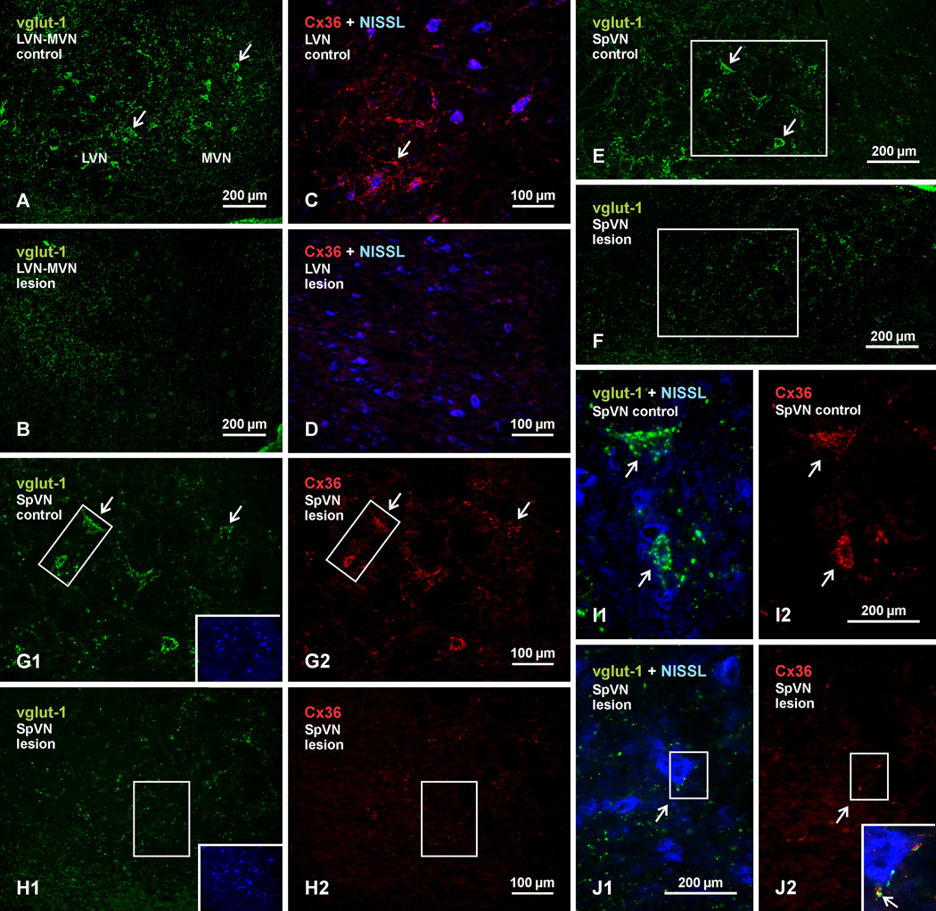
Fig. 11.
Immunofluorescence labelling of vglut-1 and Cx36 in the vestibular nuclei of adult rat after unilateral vestibular nerve section and Scarpa ganglionectomy. Pairs of images display control (left side) vs. deafferented (right side) vestibular nuclei, and each pair was acquired from areas ipsilateral and contralateral to the side of the lesion from the same animal. (A,B) Images of the LVN and the lateral portion of MVN, showing dense labelling of vglut-1 around neuronal somata on the control side (A, arrows) and a loss of this labelling around somata on the lesioned side. (C,D) Higher magnification of the LVN with blue Nissl counterstaining, showing dense collections of Cx36-puncta on the control side (C, arrow) and depletion of these puncta on the lesioned side (D). (E,F) Images showing labelling of vglut-1 associated with neurons in the SpVN on the control side (E, arrows) and a loss of this labelling on the lesion side (F). (G,H) Images of labelling for vglut-1 (G1) and Cx36 (G2) in the same field of SpVN taken from the boxed area in E, and of vglut-1 (H1) and Cx36 (H2) in the same field taken from the boxed area in F. Insets show the two fields with blue Nissl counterstaining of neurons. Vglut-1 and Cx36 associated with neuronal somata on the control side (G1,G2, arrows) is depleted on the lesion side H1,H2). (I,J) Magnifications of the boxed areas from G1 and G2 are shown in I1 and I2, respectively, and those from H1 and H2 are shown in J1 and J2, respectively. Labelling of large vglut-1-positive terminals associated with neuronal somata (blue Nissl counterstained) on the control side (I1, arrows) are absent or reduced in size on the lesion side (J1, arrow), and Cx36-puncta associated with these terminals (I2, arrows) are similarly depleted or reduced in number (J2, arrow). The few shrunken terminals left on the lesion side remain co-localized with Cx36, as shown by overlay of the boxed areas in J1 and J2 (shown in inset in J2, with red/green overlay seen a yellow, arrow).

