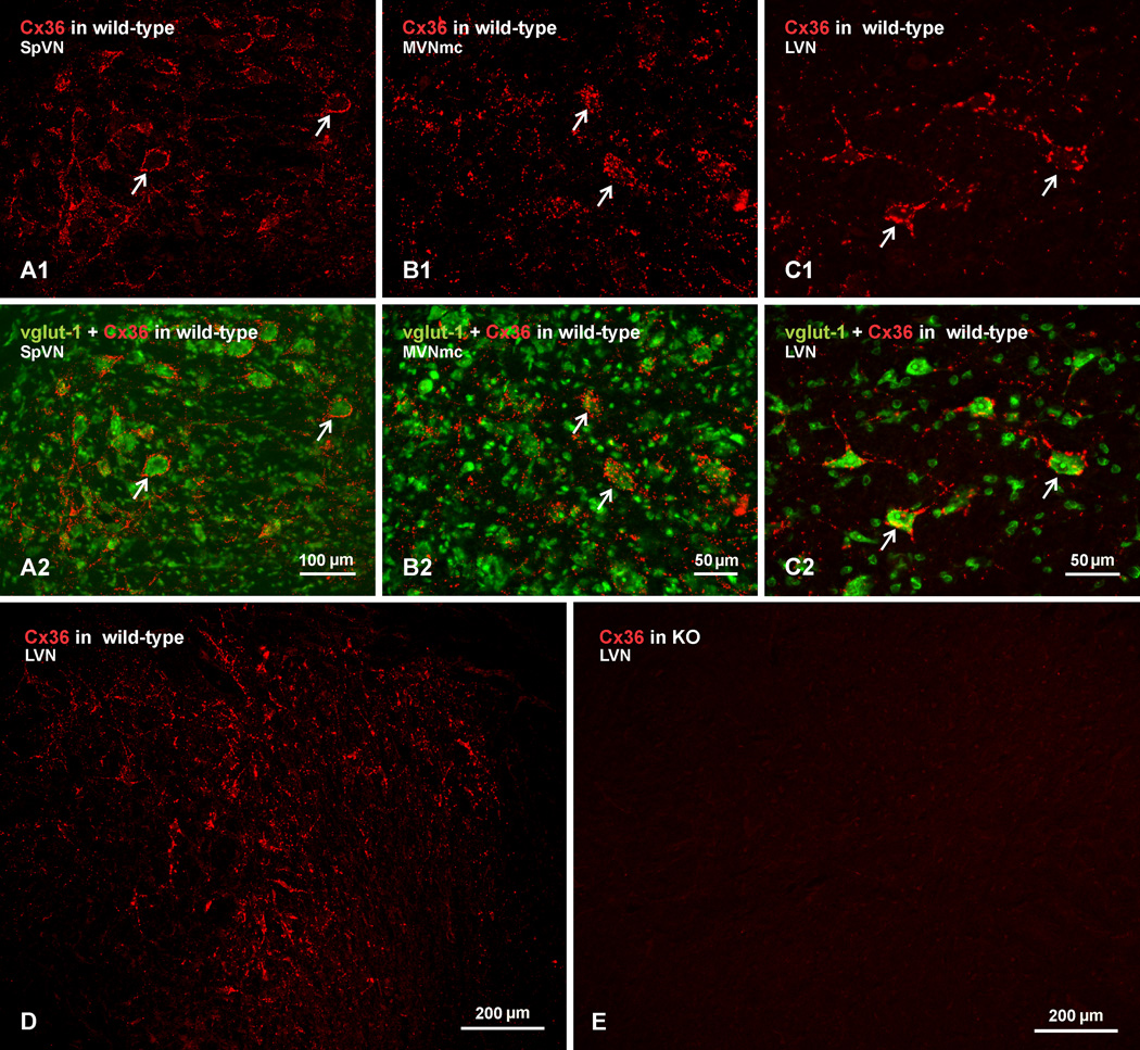
Fig. 3.
Immunofluorescence labelling of Cx36 in vestibular nuclei of wild-type adult mouse and absence of labelling in Cx36 knockout mouse. (A–C) The SpVN (A), MVNmc (B) and LVN (C), showing intense labelling of Cx36 (A1, B1 and C1, arrows) around medium and large size neuronal cell bodies, as seen in overlay with Nissl fluorescence counterstaining (green) of the corresponding fields (A2, B2, and C2, arrows). (D,E) Low magnification images showing Cx36 immunofluorescence in LVN of a wild-type mouse (D) and as similar field showing absence of labelling for Cx36 in the LVN of a Cx36 knockout mouse (E).

