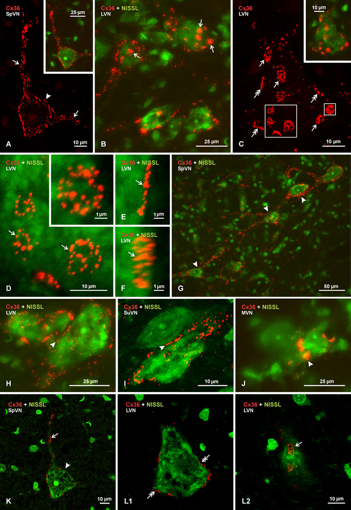
Fig. 4.
Laser scanning confocal images of Cx36 immunofluorescence associated with neurons in the vestibular nuclei of adult rat. Images are shown with labelling of Cx36 alone (red) or with Nissl fluorescence counterstaining (green) in overlay. (A) Image of the boxed area in Fig. 2B, showing Cx36 immunofluorescence on a single neuron in the SpVN, with Nissl fluorescence overlay of the same neuron in the inset. Labelling appears punctate and is seen throughout the soma surface (arrowhead) and along two large initial dendrites (arrows). (B) Confocal image of several neuronal somata in the LVN, displaying patches of dense labelling (arrows) as well as dispersed punctate labelling for Cx36 on their surface. (C) Higher magnification of a single neuron in the LVN, with Nissl counterstaining overlay in inset, showing dense patches of labelling to consist of clusters of numerous Cx36-positive puncta (arrows). Also seen are linear arrangements of Cx36-puncta at the perimeter of neuronal somata (double arrows). (D–F) Confocal images showing individual clusters of Cx36-puncta (arrows) viewed en face (D, magnification of large boxed area in C), and in inset (D, magnification of the small box in C), with clusters consisting of different numbers and sizes of Cx36-puncta. (E,F) Image of a cluster of Cx36-puncta viewed on edge (E), and the same image (F) rotated by 30o in the horizontal axis to reveal the presence of a cluster of puncta. (G) Image of neurons in the SpVN with labelling for Cx36 along dendrites that appear to extend close to nearby neuronal somata (arrowheads). (H-J) Rare examples of Cx36-puncta (arrowheads) located between relatively small neurons in LVN (H), SuVN (I) and MVN (J). (K) Single scan image taken from Figure 4A, which was a z-stack of 13 scans, showing punctate immunofluorescence labelling of Cx36 (red) on the surface of the somata (arrowhead) and an initial dendrite (arrow) of a Nissl fluorescence counterstained neuron (green). (L) Single scan images taken from the image in Figure 4C (z-stack of 11 scans), showing a scan through the cell center with Cx36-puncta localized around the cell periphery (L1, double arrows), and a scan through the upper cell surface with Cx36-puncta localized at the cell surface (L2, arrow). The single scans show absence of intracellular labelling and localization of Cx36-puncta at the cell periphery.

