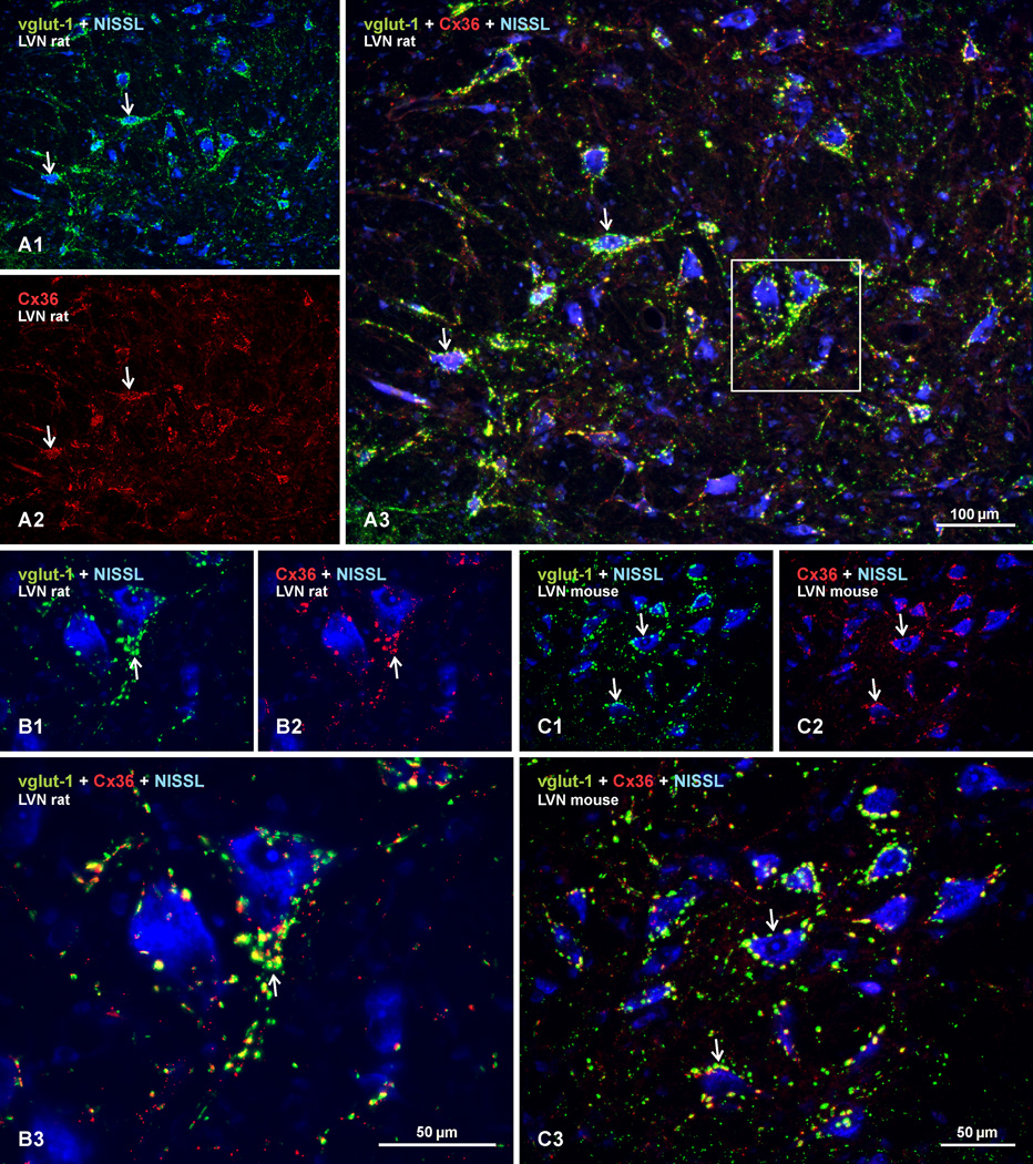
Fig. 6.
Double immunofluorescence labelling of Cx36 and vglut-1 in the LVN of adult rat and mouse. Sections were labelled for Cx36 (red), vglut-1 (green) and counterstained with Nissl fluorescence (blue). (A) Low magnification image of rat LVN, showing labelling for vglut-1 and Nissl fluorescence overlay (A1), and the same field showing labelling for Cx36 alone (A2), and overlay of all three colors (A3). Neuronal somata and their initial dendrites heavily invested with dense concentrations of vglut-1-positive nerve terminals (A1, arrows) are laden with Cx36-puncta (A2, arrows), with substantial Cx36/vglut-1 co-localization, as seen by yellow after red/green merge (A3, arrows). (B) Higher magnification of the boxed area in A3, showing vglut-1/Nissl overlay (B1), Cx36/Nissl overlay (B2), and merge of all these labels (B3), where the majority of vglut-1-positive terminals are seen to display labelling for Cx36 (B3 arrow). (C) Mouse LVN, showing similar dense labelling of vglut-1 around neuronal somata (C1, arrows), with matching pattern of labelling for Cx36 (B2, arrows) and a high degree of Cx36/vglut-1 co-localization, as seen by yellow in overlay (C3, arrows).

