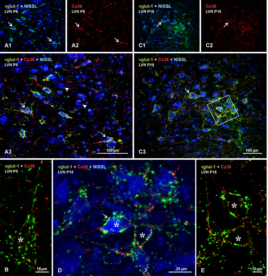
Fig. 9.
Developmental appearance of immunolabelling for Cx36 and vglut-1 in LVN of rat at postnatal day 5 and 15. Sections were labelled for Cx36 (red) and vglut-1 (green), with Nissl fluorescence counterstain (blue). (A,B) Postnatal day 5 LVN, shown at low magnification (A) and a confocal image of a single neuron (marked by asterisk) at higher magnification (B). A sparse to moderate density of vglut-1-positive terminals is seen on LVN neuronal somata (A1, arrows) and less so on dendrites, and a similar distribution of labelling for Cx36 is seen on these somata, as shown by labelling for Cx36 alone (A2, arrows), with moderate degree of co-localization, as seen in red/green/blue overlay (A3, arrows). Many neurons are nearly devoid of Cx36/vglut-1 labelling (arrowheads), and some dispersed Cx36-puncta appear unassociated with vglut-1 (B) or with Nissl-stained neurons (A3). (C–E) Postnatal day 15 LVN at low magnification (C), with boxed area in C3 shown as higher magnification confocal image in D, and the boxed area in D shown with only red/green overlay in E. Labelling for vglut-1 (C1) and Cx36 (C2) is increased compared to postnatal day 5, but is still only moderate on neuronal somata and dendritic initial segments (C3, arrows). Labelling for Cx36 appears on cell bodies and dendrites often as individual puncta rather than as clusters, dispersed Cx36-puncta not associated with Nissl-stained neurons is reduced (D,E), and degree of Cx36/vglut-1 co-localization (arrow) is modest compared with that seen in adult.

