
Figure 6. RSV inhibits TGFβ production from tBregs.
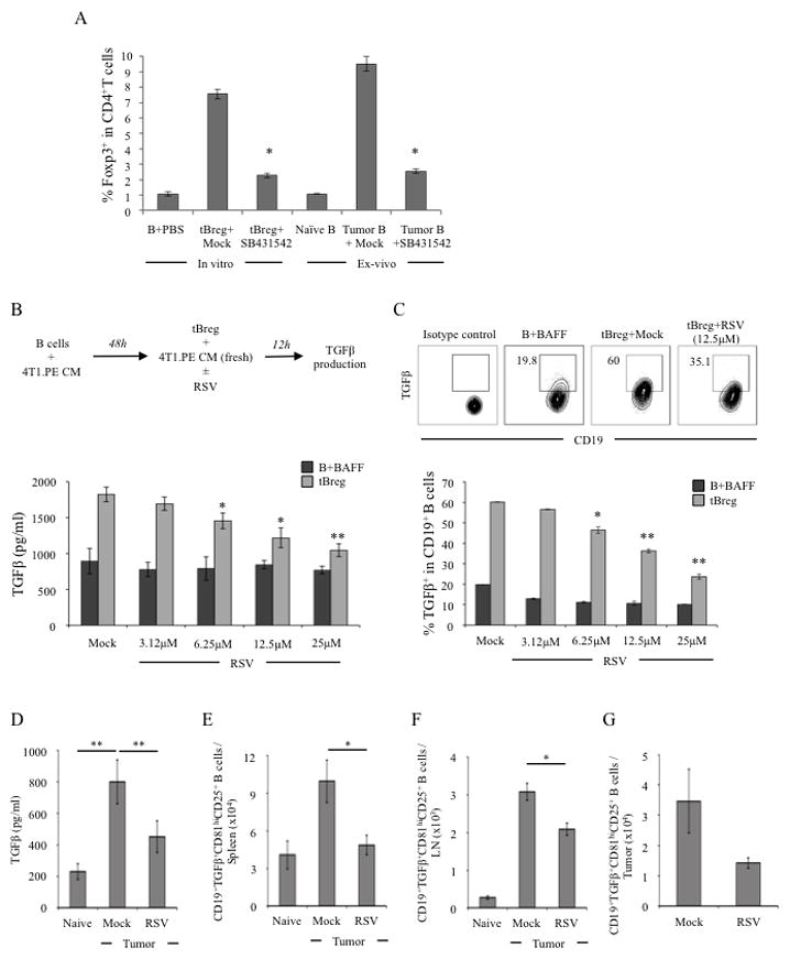
For in vitro conversion assay of Tregs (A), CD25−CD4+ T cells from naïve BALB/c mice were incubated with B cells treated with PBS, or in vitro generated tBregs, or B cells isolated from naïve or tumor (4T1.2)-bearing BALB/c mice in the presence or absence of 10 μM SB431542. T cells were stimulated with anti-CD3 Ab and 500 U/ml IL-2 for 5 days and FoxP3 expression was detected by intracellular stating (Y-axis, % of FoxP3 in CD4+ T cells ± SEM of a triplicate experiment). As shown in schema (upper panel, B), RSV was added after the generation of tBregs. Control B cells were BAFF-treated B cells (B+BAFF) incubated with RSV (lower panel, B, C). X-axis shows concentration of RSV used, and Y-axis is for levels of secreted TGFβ1 ± SEM (pg/ml, lower panel, B) and % ± SEM of intracellular TGFβ1 expression in B cells (lower panel, C) after 12 h incubation. Dot plot in C (upper panel) is representative data of triplicate experiments shown in lower panel (C) repeated three times. To assess in vivo effects of RSV, BALB/c mice were treated with RSV (50 μg) or mock every other day staring 3 days after challenge with 4T1.2 cancer cells (D). At day 10, B cells were isolated from LN tested for ability to secrete TGFβ by ELISA after 48 h stimulation with PMA/ionomycin (D). Similarly, numbers of TGFβ-expressing tBregs (CD81High CD25+ CD19+ B cells) were assessed via intracellular staining in freshly isolated B cells from spleen (E), draining LN (F) and tumor (G). Shown, representative data of 4 mice per group experiments repeated three times.