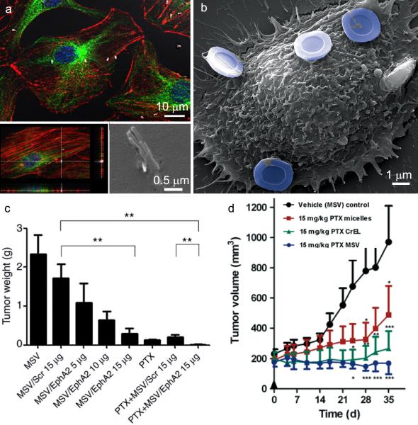
Figure 3. In vitro cellular association of pSi particles with endothelial cells and in vivo therapeutic efficacy of multistage vector delivery of EphA2 siRNA and paclitaxel.
A. Confocal (single plane and z-stacked) and an electron micrograph show 2 μm pSi nanowires located within or in the process of being internalized by endothelial cells. Cells in the confocal images are labeled with nuclear (blue) and cytoskeleton (red actin and green microtubules) dyes. B. The scanning electron micrograph shows an endothelial cell with 3 μm hemispherical pSi particles associated with the cell surface. Particles are pseudo-colored in blue. Reproduced by permission of The Royal Society of Chemistry, Nanoscale, Serda et al.[50] C. Tumor weight 6 weeks after initiation of biweekly multistage vector (MSV) /EphA2 siRNA injections (intravascular) in combination with paxlitaxel (PTX; weekly; intraperitoneal) or monotherapy in a mouse model of ovarian cancer (2 mg/kg PTX; scr = scrambled siRNA; **p<0.01). Reprinted with permission from the American Association of Cancer Research, Clinical Cancer Research, Shen H, et al. [33]. D. Antitumor efficacy of MSV loaded with PTX in female nude mice bearing MDA-MB-468 breast cancer xenografts. Treatment groups included: (i) control micelles; (ii) PTX micelles; (iii) PTX Cremophor EL (CrEL, a polyethoxylated castor oil formulation); and (iv) PTX MSV (all were treated with a single dose at Day 0; PTX 15 mg/kg). Asterisks denote results in which the difference was statistically significant compared to the control group (*p<0.05; **p<0.01; ***p<0.001). Reproduced with permission from Elsevier, Cancer Letters, Blanco E, et al. [35].

