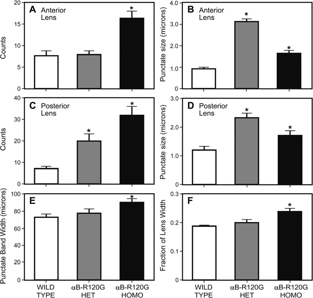
Figure 2.
Quantification of p62 speckles in wild type and αB-R120G mutant lenses. (A) Number of p62 speckles in anterior lens fiber cells. Note the increase in the number of p62-positive puncta in the αB-R120G homozygous lenses. n=5. Asterisks indicate statistical significance (WT vs. Homozygous, p<0.0001). (B) Size of p62-positive puncta in anterior lens fiber cells. Note the increase in size of the puncta in αB-R120G homozygous and heterozygous mutant lenses compared with the wild type lenses (WT vs. Heterozygous p< 0.0001; WT vs. Homozygous p<0.05). (C) Number of p62 speckles in posterior lens fiber cells. Note the increase in the number of p62-positive puncta in the αB-R120G homozygous lenses (WT vs. Heterozygous p<0.05; WT vs. Homozygous p<0.05). (D) Size of p62-positive puncta in posterior lens fiber cells. Note the increase in size of the puncta in αB-R120G homozygous lenses compared with the wild type lenses. (E) Width of punctate/speckles band in wild type and αB-R120G mutant lenses (WT vs. Homozygous p<0.05). (F) Fraction of the lens equatorial diameter in wild type, αB-R120G homozygous, and αB-R120G heterozygous lenses (WT vs. Homozygous p<0.05).

