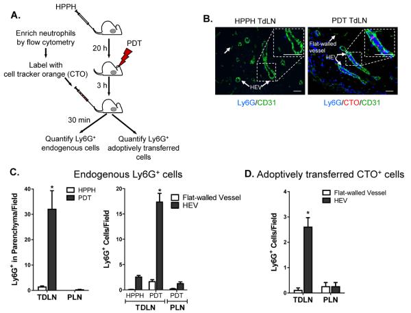
Figure 3. TDLN HEVs support increased neutrophil accumulation following PDT treatment.
(A) A single-cell suspension of neutrophils was fluorescently labeled with CellTracker Orange CMTMR (red) and adoptively transferred into treated mice as shown in the schematic. (B) Lymph node tissue sections were stained with anti-CD31 (green) and anti-Ly6G (blue). Endogenous (blue) and adoptively transferred Ly6G+ cells (pink) were counted following short-term (0 – 30 minute) homing assays. Representative photomicrographs of control (HPPH only) and PDT treated lymph nodes are shown. Quantification of endogenous Ly6G+ cells and adoptively transferred Ly6G+ neutrophils is presented in (C) and (D), respectively. Serial sections were stained to confirm that CD31-expressing cuboidal cells also expressed the HEV-specific marker PNAd. Data (mean ± SEM) are of ≥ 9 fields, unless the size of the TDLN restricted the number of fields, in individual mice and are representative of 3 independent experiments. *P<0.05 Scale bars represent 50 μm; parenchyma, TDLN stroma; PLN, peripheral LN (inguinal and popliteal).

