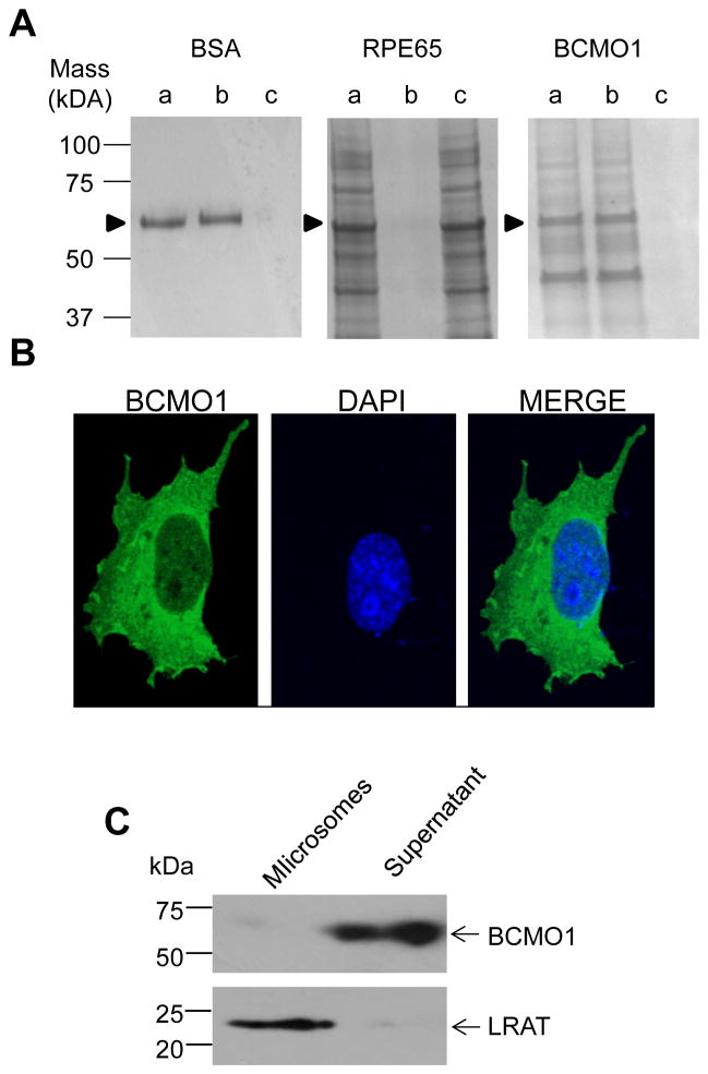
Figure 6. Partitioning of BCMO1 in Triton X-114 phase separations and BCMO1 solubility in tissues and cells.
A, Pure BSA, RPE65 microsomes, and Talon-purified recombinant BCMO1 were subjected to phase separation experiments as described under “Materials and Methods”. These experiments show that BCMO1 partitions into the aqueous phase similar to BSA and contrary to RPE65 which partitions into the detergent phase, as shown in previously reported studies [33, 47]. Lanes: a, Input, indicates the pure proteins and RPE65 microsomes before phase separation, b, the aqueous phase, and c, the detergent phase. Arrowheads indicate the positions of corresponding proteins as labeled on top of each Coomassie stained gel. B, Immunostaining of human BCMO1 (green) in Cos7 cells. The nucleus is stained with DAPI (blue). The merged image shows that BCMO1 is located in the cytoplasm. C, Immunoblot analyses for BCMO1 and LRAT in cytoplasmic and microsomal preparations of mouse liver. BCMO1 was detected in the cytoplasmic fraction whereas LRAT localized to microsomes.

