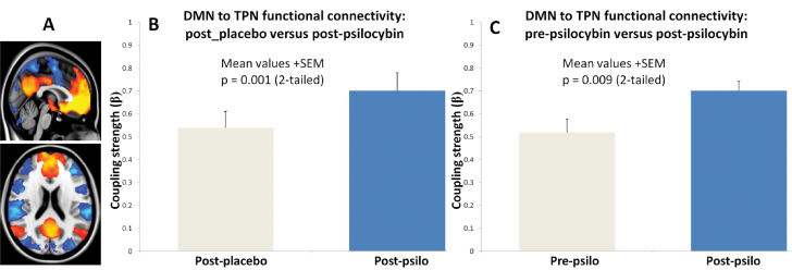
Fig. 3.
Increased functional FC between the default-mode- and task-positive network under psilocybin. (A) vmPFC-positive- (DMN, orange) and vmPFC-negative regions (TPN, blue). (B) Increased FC between the DMN and TPN after psilocybin vs placebo (P = .001). (C) Increased FC between the DMN and TPN post vs prepsilocybin (P = .009).

