
Figure 4.
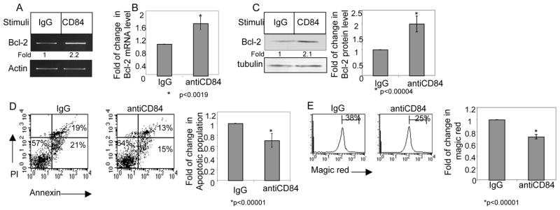
Stimulation of CD84 induces a survival cascade in CLL cells. CLL cells were incubated in the presence of agonistic anti-CD84 (1 ng/ml) or a control antibody (IgG) for 30 minutes. The cells then were washed and bound mAb was cross-linked with F(ab′)2 goat anti mouse Ig. (A) Following 18 h, the RNA was purified, and Bcl-2 and actin mRNA levels were analyzed. The results presented are representative of seven CLL patients. (B) Following 18 h, RNA was purified, and qPCR was performed using primers for Bcl-2 and RP-2. Results are expressed as fold of change in Bcl-2 mRNA in stimulated and non-stimulated cells; untreated cells were defined as 1. The graph summarizes results from four CLL patients. (C) Following 24 hr, cells were lysed and Bcl-2 and tubulin proteins were analyzed by western blot analysis. Graph summarizes the results of four different experiments. (D) Following 26h, cells were stained with Annexin V and PI, and analyzed by FACS. The graph summarizes the results of seven CLL patients. (E) Following 26h, cells were stained with Magic Red and analyzed by FACS. The graph summarizes the results of four CLL patients.