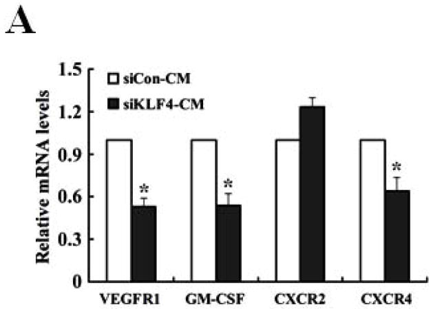
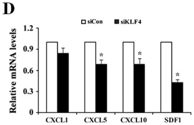
Figure 5. GM-CSF is upregulated by KLF4 through CXCL5.
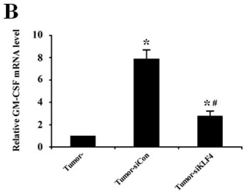
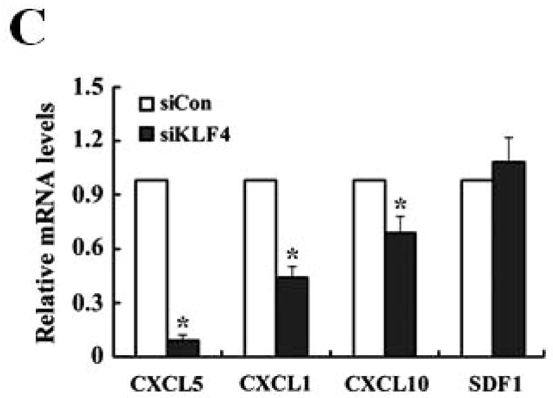
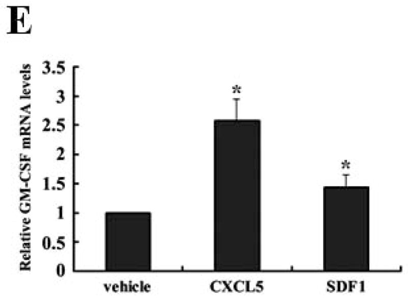
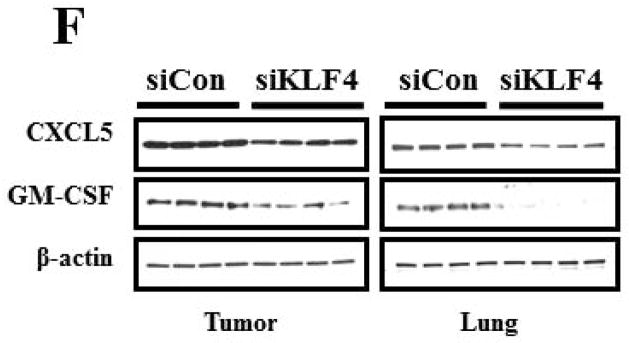
(A) Expression of VEGFR1, GM-CSF, CXCR2 and CXCR4 in MDSCs incubated with conditioned medium derived from siCon and siKLF4 cells (designated as siCon-CM and siKLF4-CM, respectively) was determined by quantitative RT-PCR. Values were expressed as mean ± SD of three independent experiments. * P < 0.05 versus siCon group. (B) GM-CSF expression in bone marrow cells was determined by quantitative RT-PCR upon incubation of 4T1 tumor tissues. 50 mg siCon and siKLF4 4T1 tumor tissues (Tumor-siCon and Tumor-siKLF4) were incubated with 1 × 10⁶ bone marrow cells for 24 h followed by real time RT-PCR analysis. Bone marrow cells without tumor incubation (Tumor-) were used as a control. Values were expressed as means ± SD of three independent experiments. * P < 0.05 versus Tumor- group. # indicates P < 0.05 versus Tumor-siCon group. (C) Chemokine expression in mammary tumor tissues from control (siCon) and KLF4 knockdown (siKLF4) 4T1 cell-inoculated mice was determined by quantitative RT-PCR. Values were expressed as means ± SE of three independent experiments. * P < 0.05 versus siCon group. (D) Chemokine expression in 4T-1 control (siCon) and KLF4 knockdown (siKLF4) stable cells was determined by quantitative RT-PCR. Values were expressed as means ± SD of three independent experiments. * P < 0.05 vs siCon group. (E) GM-CSF expression was determined by quantitative RT-PCR after the bone marrow cells from BALB/c mice were treated with CXCL5 (25 ng/ml) and SDF1 (50 ng/ml) for 3 hrs. * P < 0.05 versus vehicle group. (F) Decreased expression of CXCL5 and GM-CSF in tumor and lung tissues of siCon and siKLF4 cell-inoculated mice by Western blotting. β-actin was used as an internal control. Results presented were from four different mice.

