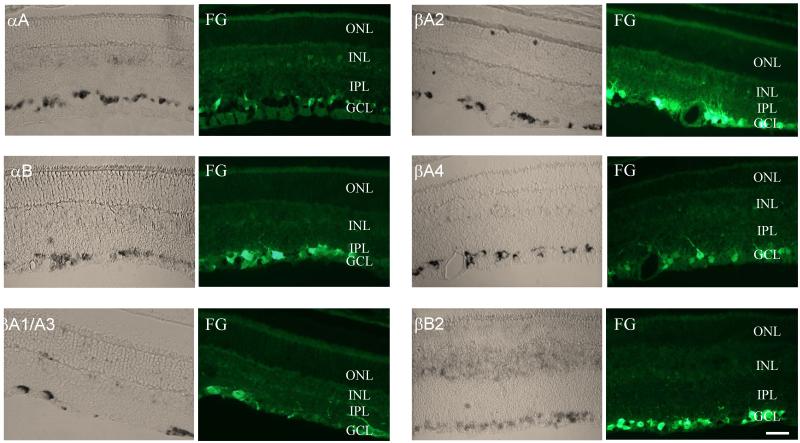
Figure 1.
In situ analysis of the crystallin expression in the retina. The expression of crystallins was primarily observed in the ganglion cell layer (GCL). Relatively weak staining can also be seen in the inner nuclear layer (INL) and, to a much lesser degree, in the outer nuclear layer (ONL). Crystallin-positive cells in the GCL were colocalized with RGCs retrogradely labeled with Fluorogold (FG). IPL, inner plexiform layer. Scale bars =20 μm.

